File:LiverSceneViewSaggital.png
From NAMIC Wiki
Size of this preview: 800 × 508 pixels. Other resolutions: 320 × 203 pixels | 985 × 625 pixels.
Original file (985 × 625 pixels, file size: 143 KB, MIME type: image/png)
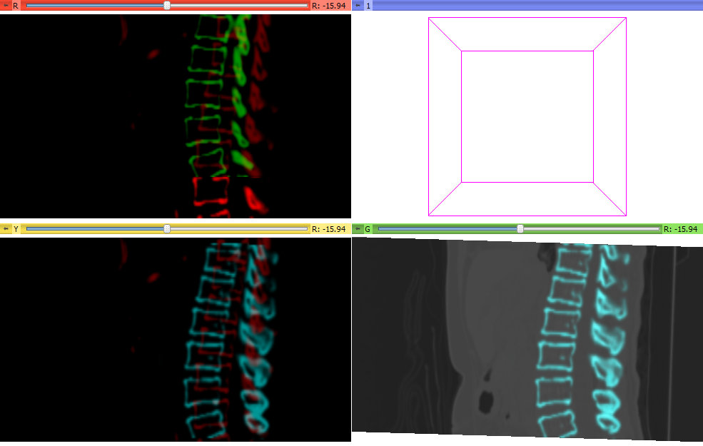
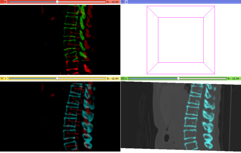
Bones near liver are segmented for two time point images of a liver. Time point A is red and B is green. The moving green image is registered on the red fixed image. The green image is transformed to the cyan image. The same transformation matrix on the cyan bone image is used to transformed the moving liver data set B.
File history
Click on a date/time to view the file as it appeared at that time.
| Date/Time | Thumbnail | Dimensions | User | Comment | |
|---|---|---|---|---|---|
| current | 23:50, 12 January 2012 | | 985 × 625 (143 KB) | Ktdiedrich (talk | contribs) | Bones near liver are segmented for two time point images of a liver. Time point A is red and B is green. The moving green image is registered on the red fixed image. The green image is transformed to the cyan image. The same transformation matrix on the c |
- You cannot overwrite this file.
File usage
The following page uses this file: