File:Stroke lesion mixture model.jpg
From NAMIC Wiki
Size of this preview: 800 × 600 pixels. Other resolutions: 320 × 240 pixels | 1,200 × 900 pixels.
Original file (1,200 × 900 pixels, file size: 160 KB, MIME type: image/jpeg)
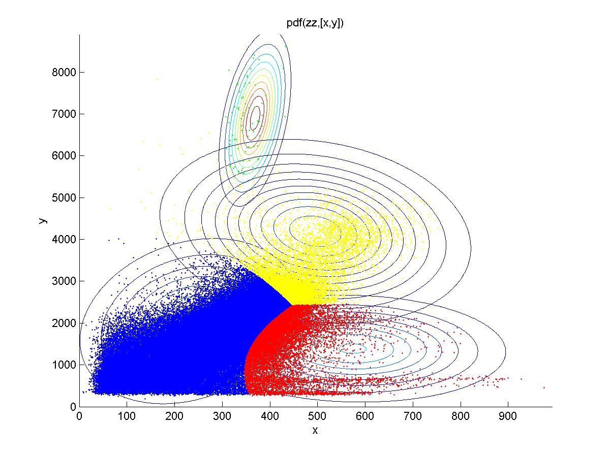
Different tissue classes found using Gaussian mixture models for FLAIR (x) and DWI (y). Yellow corresponds to (oversegmented) stroke lesion, blue corresponds to normal tissue, and red and green are artifacts and white matter hyperintensity respectively.
File history
Click on a date/time to view the file as it appeared at that time.
| Date/Time | Thumbnail | Dimensions | User | Comment | |
|---|---|---|---|---|---|
| current | 00:55, 10 January 2014 | | 1,200 × 900 (160 KB) | Rameshvs (talk | contribs) | Different tissue classes found using Gaussian mixture models for FLAIR (x) and DWI (y). Yellow corresponds to (oversegmented) stroke lesion, blue corresponds to normal tissue, and red and green are artifacts and white matter hyperintensity respectively. |
- You cannot overwrite this file.
File usage
The following page uses this file: