Difference between revisions of "2010 Winter Project Week Spine Segmentation Module in Slicer3"
Sylvainjaume (talk | contribs) (revise captions for the top screenshots) |
Sylvainjaume (talk | contribs) (balance the text in the caption and in the approach description) |
||
| Line 4: | Line 4: | ||
Image:spine_segmentation_module_in_slicer_002.png|The cause of back pain is the pressure of the herniated disc onto the spinal nerves. This Slicer rendering shows an herniated disc pushing the canal where the cerebro-spinal fluid (CSF) needs to insulate the nerves from outside shocks. | Image:spine_segmentation_module_in_slicer_002.png|The cause of back pain is the pressure of the herniated disc onto the spinal nerves. This Slicer rendering shows an herniated disc pushing the canal where the cerebro-spinal fluid (CSF) needs to insulate the nerves from outside shocks. | ||
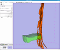
Image:spine_segmentation_in_slicer_004.png|Surface meshes and volumetric meshes of our automatically created model of the CSF. The paths where the nerves exit the spine appear as two roots descending on each side of the spinal column. | Image:spine_segmentation_in_slicer_004.png|Surface meshes and volumetric meshes of our automatically created model of the CSF. The paths where the nerves exit the spine appear as two roots descending on each side of the spinal column. | ||
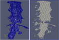
| − | Image:Jaume_Loepprich_vertebrae_3D_rendering.png|A complex model of a lumbar vertebrae has been created to drive our shape-based segmentation algorithm in a robust and accurate recognition | + | Image:Jaume_Loepprich_vertebrae_3D_rendering.png|A complex model of a lumbar vertebrae has been created to drive our shape-based segmentation algorithm in a robust and accurate recognition. |
</gallery> | </gallery> | ||
| Line 37: | Line 37: | ||
<h3>Progress</h3> | <h3>Progress</h3> | ||
The algorithm has been implemented in Slicer 3.5 as an extension module. | The algorithm has been implemented in Slicer 3.5 as an extension module. | ||
| − | + | This volumetric pattern recognition algorithm is fully implemented as a Slicer module using ITK and VTK. | |
| − | + | The module has been tested on data sets acquired at Brigham and Women's Hospital using the Wideband Steady-State Free-Precession (WB-SSFP) MRI protocol. | |
| − | The module has been tested on data sets acquired at Brigham and Women's Hospital using the Wideband Steady-State Free-Precession (WB-SSFP) MRI protocol | ||
| − | |||
</div> | </div> | ||
</div> | </div> | ||
Revision as of 06:11, 21 January 2010
Home < 2010 Winter Project Week Spine Segmentation Module in Slicer3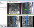
Our goal is to develop a Slicer module to automatically segment the spine in 3D MRI images to help during the surgical removal of herniated discs. (See a video)
The cause of back pain is the pressure of the herniated disc onto the spinal nerves. This Slicer rendering shows an herniated disc pushing the canal where the cerebro-spinal fluid (CSF) needs to insulate the nerves from outside shocks.
Surface meshes and volumetric meshes of our automatically created model of the CSF. The paths where the nerves exit the spine appear as two roots descending on each side of the spinal column.
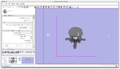
A complex model of a lumbar vertebrae has been created to drive our shape-based segmentation algorithm in a robust and accurate recognition.


Key Investigators
- Sylvain Jaume (MIT)
- Martin Loepprich (University of Heidelberg)
- Ron Kikinis, Steve Pieper (BWH)
- Polina Golland (MIT)
Objective
We are developing a Slicer module to segment the region within the thecal sac in MRI images of the spine. Our objective is to provide a segmentation and visualization tool to improve the treatment of disc herniation. The structures of interests are the cerebro-spinal fluid (CSF), the discs, the vertebrae and the spinal nerves. The main challenge is to perform the segmentation in a fully automated way.
Approach, Plan
Our plan for the project week is to integrate our code into Slicer 3.5. Our code analyzes the intensity profile of different regions in the MRI and automatically defines the optimum region for the CSF.
Progress
The algorithm has been implemented in Slicer 3.5 as an extension module. This volumetric pattern recognition algorithm is fully implemented as a Slicer module using ITK and VTK. The module has been tested on data sets acquired at Brigham and Women's Hospital using the Wideband Steady-State Free-Precession (WB-SSFP) MRI protocol.


