Difference between revisions of "2017 Winter Project Week/Multi-ModalitySegmentationOfUSandMRImagesForGliomaSurgery"
From NAMIC Wiki
| Line 19: | Line 19: | ||
| | | | ||
<!-- Objective bullet points --> | <!-- Objective bullet points --> | ||
| − | * Multi-Modual Image Segmentation of preoperative MR- and intraoperative Ultrasound(US)-images | + | * Multi-Modual Image Segmentation of preoperative MR- and intraoperative Ultrasound(US)-images for multi-modual image registration. |
* Segmentation of the following anatomical structures: Falx cerebri, tentorium cerebelli, white matter, gray matter, CSF (ventricles), blood vessels. | * Segmentation of the following anatomical structures: Falx cerebri, tentorium cerebelli, white matter, gray matter, CSF (ventricles), blood vessels. | ||
* Testing applicability of Deep Learing on current data | * Testing applicability of Deep Learing on current data | ||
Revision as of 18:12, 9 January 2017
Home < 2017 Winter Project Week < Multi-ModalitySegmentationOfUSandMRImagesForGliomaSurgery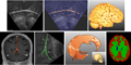
Several structures of the brain segmented from US- and MR images.

Key Investigators
- Jennifer Nitsch, University of Bremen (Germany)
Project Description
| Objective | Approach and Plan | Progress and Next Steps |
|---|---|---|
|
|
|