Difference between revisions of "2009 Summer Project Week Liver Ablation Slicer"
From NAMIC Wiki
| (10 intermediate revisions by 3 users not shown) | |||
| Line 1: | Line 1: | ||
__NOTOC__ | __NOTOC__ | ||
<gallery> | <gallery> | ||
| − | Image:PW2009-v3.png|[[2009_Summer_Project_Week| | + | Image:PW2009-v3.png|[[2009_Summer_Project_Week#Projects|Projects List]] |
| + | Image:originalSegmentation.png|Segmentation showing no pass zones (ribs)and tumor. | ||
| + | Image:segmentationAfterDilation.png|Segmented tumor region is dilated according to physician prescribed ablation margin. | ||
| + | Image:Slicer_liver_module.png|IGT planning module for liver albation in Slicer3. | ||
| + | Image:Segmentation_dilated_by_slicer.png|Segmented tumor region is dilated in Slicer3. | ||
</gallery> | </gallery> | ||
| Line 12: | Line 16: | ||
<h3>Objective</h3> | <h3>Objective</h3> | ||
| − | + | Implement a complete workflow: | |
| + | #Load data. | ||
| + | #Manually mark regions (tumor, entry, critical structure). | ||
| + | #Process segmentation and export information to planning module (executable plugin with command line options). | ||
| + | #Load results of the optimization program. | ||
| + | #Configure OpenIGTLink module on Slicer and run OpenIGTLink IGSTK client. | ||
| + | #Navigate. | ||
</div> | </div> | ||
| Line 18: | Line 28: | ||
<h3>Approach, Plan</h3> | <h3>Approach, Plan</h3> | ||
| − | + | #Integrate code from Georgetown for step 3 into Slicer. | |
| + | #Decide on appropriate format for describing the output of the optimization (set of trajectories and ablations along each trajectory). | ||
| + | #Implement a stub executable plugin as a stand in for the optimization program. | ||
| + | #Test the integrated workflow. | ||
</div> | </div> | ||
| Line 24: | Line 37: | ||
<h3>Progress</h3> | <h3>Progress</h3> | ||
| − | + | * Completed GUI design and implementation for "planning" part of the workflow. | |
| + | * An unique feature in the planning part were tumor segmentation and ablation volume planning, taking ablation magin into account. | ||
| + | * "Navigation" part 80% done. | ||
| + | * In "Navigation", "IGT Guidance" widget was designed and implemented to be shared with other IGT investigators. | ||
| + | |||
</div> | </div> | ||
| + | </div> | ||
| + | |||
| + | <div style="width: 97%; float: left;"> | ||
| + | |||
| + | ==References== | ||
| + | *Z. Yaniv, E. Wilson, D. Lindisch, K. Cleary, "Electromagnetic Tracking in the Clinical Environment", Med. Phys., Vol. 36(3), pp. 876-892, 2009. PMID: 19378748 | ||
| + | *J. Tokuda et al., "OpenIGTLink: An open network protocol for image- guided therapy environment," International Journal of Medical Robotics and Computer Assisted Surgery, to appear. | ||
| + | |||
</div> | </div> | ||
Latest revision as of 15:16, 26 June 2009
Home < 2009 Summer Project Week Liver Ablation Slicer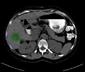
Segmentation showing no pass zones (ribs)and tumor.
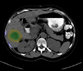
Segmented tumor region is dilated according to physician prescribed ablation margin.
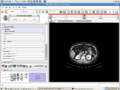
IGT planning module for liver albation in Slicer3.
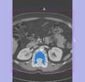
Segmented tumor region is dilated in Slicer3.


Key Investigators
- BWH: Haiying Liu, Noby Hata
- Georgetown: Ziv Yaniv
Objective
Implement a complete workflow:
- Load data.
- Manually mark regions (tumor, entry, critical structure).
- Process segmentation and export information to planning module (executable plugin with command line options).
- Load results of the optimization program.
- Configure OpenIGTLink module on Slicer and run OpenIGTLink IGSTK client.
- Navigate.
Approach, Plan
- Integrate code from Georgetown for step 3 into Slicer.
- Decide on appropriate format for describing the output of the optimization (set of trajectories and ablations along each trajectory).
- Implement a stub executable plugin as a stand in for the optimization program.
- Test the integrated workflow.
Progress
- Completed GUI design and implementation for "planning" part of the workflow.
- An unique feature in the planning part were tumor segmentation and ablation volume planning, taking ablation magin into account.
- "Navigation" part 80% done.
- In "Navigation", "IGT Guidance" widget was designed and implemented to be shared with other IGT investigators.
References
- Z. Yaniv, E. Wilson, D. Lindisch, K. Cleary, "Electromagnetic Tracking in the Clinical Environment", Med. Phys., Vol. 36(3), pp. 876-892, 2009. PMID: 19378748
- J. Tokuda et al., "OpenIGTLink: An open network protocol for image- guided therapy environment," International Journal of Medical Robotics and Computer Assisted Surgery, to appear.