Difference between revisions of "2010 Summer Project Week Liver Ablation"
| Line 10: | Line 10: | ||
* BWH: Haiying Liu, Noby Hata, Sota Oguro | * BWH: Haiying Liu, Noby Hata, Sota Oguro | ||
* Georgetown University: Ziv Yaniv | * Georgetown University: Ziv Yaniv | ||
| − | |||
| − | |||
| − | |||
| − | |||
| − | |||
| − | |||
| − | |||
| − | |||
| − | |||
| − | |||
| − | |||
| − | |||
<h3>Objectives</h3> | <h3>Objectives</h3> | ||
Revision as of 15:47, 17 June 2010
Home < 2010 Summer Project Week Liver Ablation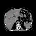
Liver with fiducials
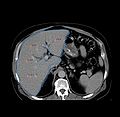
Liver with fiducials and segmentation
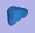
Liver model


Key Investigators
- BWH: Haiying Liu, Noby Hata, Sota Oguro
- Georgetown University: Ziv Yaniv
Objectives
- denoising and bias field correction of 3D US data
- model for the combined image correction, segmentation and registration between 3D US and MRI
- visualization of all processing steps within Slicer
Approach, Plan
The bias field on the 3D US data varies spatially with a very low frequency. It is similar to the bias field effects in MR caused by magnetic inhomogeneities. We will investigate whether an EM-based approach may be used for the correction, similar to the previous work by Wells et. al. and Pohl et. al., and try to adapt the already existing Slicer code to this problem.
For the registration, we may have to come up with a new formulation that uses not only the statistics of the intensities, but also some additional information about the tissue boundaries. The tissue boundaries are directly influencing the visible content of the 3D US images, however, they have a different appearance and structure within the MRI.
Progress
Fill this at the end of the week
Delivery Mechanism
The work will be provided as ITK modules and it intended to integrate them into a Slicer module.
References
- W.M. Wells, R. Kikinis, W.E.L. Grimson, F. Jolesz. Adaptive segmentation of MRI data. IEEE Transactions on Medical Imaging, 15, pp. 429-442, 1996
- K.M. Pohl, J. Fisher, W.E.L. Grimson, R. Kikinis, and W.M. Wells. A Bayesian model for joint segmentation and registration. NeuroImage, 31(1), pp. 228-239, 2006
- E.N.K. Kollorz, D.A. Hahn, R. Linke, T.W. Goecke, J. Hornegger, and T. Kuwert. Quantification of Thyroid Volume using 3-D Ultrasound Imaging. IEEE Transactions on Medical Imaging, 27(4), pp. 457-466, 2008
- W. Wein, S. Brunke, A. Khamene, M.R. Callstrom, N. Navab. Automatic CT-Ultrasound Registration for Diagnostic Imaging and Image-guided Intervention. Medical Image Analysis, 12(5), pp. 577-585, 2008