Difference between revisions of "2010 Winter Project Week Cardiac Ablation Scar Segmentation"
| (16 intermediate revisions by 2 users not shown) | |||
| Line 2: | Line 2: | ||
<gallery> | <gallery> | ||
Image:PW-SLC2010.png|[[2010_Winter_Project_Week#Projects|Projects List]] | Image:PW-SLC2010.png|[[2010_Winter_Project_Week#Projects|Projects List]] | ||
| − | Image: | + | Image:Mdepa_MRA_seg.png|Left atrium (LA) segmentation in blood pool image |
| − | Image: | + | Image:Mdepa_MRA_seg_3D.png|3D model of LA segmentation |
| + | Image:Mdepa_MDE.png|Corresponding delayed enhancement image | ||
| + | Image:Mdepa_MRA_MDE_registration.png|Registered blood pool and delayed enhancement images | ||
| + | Image:Mdepa_MDE_la_segmentation.png|LA segmentation transferred to delayed enhancement image | ||
| + | Image:Mdepa_MDE_scar_segmentation.png|Ablation scar segmentation in delayed enhancement image | ||
| + | Image:Mdepa_MDE_scar_seg_3D.png|3D model of (left) our ablation scar segmentation and (right) expert segmentation | ||
</gallery> | </gallery> | ||
| Line 9: | Line 14: | ||
==Key Investigators== | ==Key Investigators== | ||
* MIT: Michal Depa, Polina Golland | * MIT: Michal Depa, Polina Golland | ||
| − | * BWH: Ron Kikinis | + | * BWH: Ehud Schmidt, Ron Kikinis |
<div style="margin: 20px;"> | <div style="margin: 20px;"> | ||
| Line 15: | Line 20: | ||
<h3>Objective</h3> | <h3>Objective</h3> | ||
| − | Atrial fibrillation | + | Atrial fibrillation is one of the most common heart conditions and can have very serious consequences such as stroke and heart failure. A technique called catheter radio-frequency (RF) ablation has recently emerged as a treatment. It involves burning the cardiac tissue that is responsible for the fibrillation. Even though this technique has been shown to work fairly well on atrial fibrillation patients, repeat procedures are often needed to fully correct the condition because surgeons lack the necessary tools to quickly evaluate the success of the procedure. |
The objective of this project is to automatically segment the scar created by RF ablation in delayed enhancement MR images acquired after the procedure. We would then be able to present the surgeon with a visualization showing the size, shape and location of the scar. | The objective of this project is to automatically segment the scar created by RF ablation in delayed enhancement MR images acquired after the procedure. We would then be able to present the surgeon with a visualization showing the size, shape and location of the scar. | ||
| Line 26: | Line 31: | ||
Our earlier work focused on segmenting the left atrium (LA), the heart chamber on which RF ablations are usually done, in blood pool MR images. We used a label fusion segmentation algorithm which first registered all of the training images to the test one and then employed a weighted voting procedure at each voxel. | Our earlier work focused on segmenting the left atrium (LA), the heart chamber on which RF ablations are usually done, in blood pool MR images. We used a label fusion segmentation algorithm which first registered all of the training images to the test one and then employed a weighted voting procedure at each voxel. | ||
| − | Given corresponding cardiac blood pool and post-procedure delayed enhancement images for each patient, our plan is to first segment the LA in the blood pool image, then transfer this segmentation to the delayed enhancement image of the same patient. We intend to use this prior information while searching for the ablation scar using intensity | + | Given corresponding cardiac blood pool and post-procedure delayed enhancement images for each patient, our plan is to first segment the LA in the blood pool image, then transfer this segmentation to the delayed enhancement image of the same patient. We intend to use this prior information while searching for the ablation scar using intensity based algorithms. This prior knowledge of the LA location will allow us to avoid most false positives. |
</div> | </div> | ||
| Line 33: | Line 38: | ||
<h3>Progress</h3> | <h3>Progress</h3> | ||
| − | + | * Registered corresponding blood pool and delayed enhancement images using a rigid registration algorithm. | |
| + | |||
| + | * Transferred LA segmentation from blood pool to delayed enhancement image using this registration result. | ||
| + | |||
| + | * Performed ablation scar segmentation experiments by using the LA segmentation as a spatial prior to limit the search space. Although we attempted several methods, the best results were obtained by finding a threshold that most separated the intensity histograms of normal LA and scar tissue in the delayed enhancement image. This gives results that are fairly close to expert segmentations. | ||
| + | In the coming weeks, we will try to improve on this method and then try to segment ablation scars in a larger set of subjects. | ||
</div> | </div> | ||
</div> | </div> | ||
Latest revision as of 18:42, 7 January 2010
Home < 2010 Winter Project Week Cardiac Ablation Scar Segmentation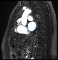
Left atrium (LA) segmentation in blood pool image
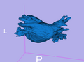
3D model of LA segmentation
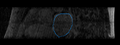
Corresponding delayed enhancement image
Registered blood pool and delayed enhancement images
LA segmentation transferred to delayed enhancement image
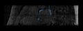
Ablation scar segmentation in delayed enhancement image
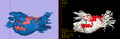
3D model of (left) our ablation scar segmentation and (right) expert segmentation


Key Investigators
- MIT: Michal Depa, Polina Golland
- BWH: Ehud Schmidt, Ron Kikinis
Objective
Atrial fibrillation is one of the most common heart conditions and can have very serious consequences such as stroke and heart failure. A technique called catheter radio-frequency (RF) ablation has recently emerged as a treatment. It involves burning the cardiac tissue that is responsible for the fibrillation. Even though this technique has been shown to work fairly well on atrial fibrillation patients, repeat procedures are often needed to fully correct the condition because surgeons lack the necessary tools to quickly evaluate the success of the procedure.
The objective of this project is to automatically segment the scar created by RF ablation in delayed enhancement MR images acquired after the procedure. We would then be able to present the surgeon with a visualization showing the size, shape and location of the scar.
Approach, Plan
Our earlier work focused on segmenting the left atrium (LA), the heart chamber on which RF ablations are usually done, in blood pool MR images. We used a label fusion segmentation algorithm which first registered all of the training images to the test one and then employed a weighted voting procedure at each voxel.
Given corresponding cardiac blood pool and post-procedure delayed enhancement images for each patient, our plan is to first segment the LA in the blood pool image, then transfer this segmentation to the delayed enhancement image of the same patient. We intend to use this prior information while searching for the ablation scar using intensity based algorithms. This prior knowledge of the LA location will allow us to avoid most false positives.
Progress
- Registered corresponding blood pool and delayed enhancement images using a rigid registration algorithm.
- Transferred LA segmentation from blood pool to delayed enhancement image using this registration result.
- Performed ablation scar segmentation experiments by using the LA segmentation as a spatial prior to limit the search space. Although we attempted several methods, the best results were obtained by finding a threshold that most separated the intensity histograms of normal LA and scar tissue in the delayed enhancement image. This gives results that are fairly close to expert segmentations.
In the coming weeks, we will try to improve on this method and then try to segment ablation scars in a larger set of subjects.