Difference between revisions of "2010 Winter Project Week Musco Skeletal Segmentation"
| Line 4: | Line 4: | ||
Image:IdealSpgrFat.jpg|Ideal Spgr Fat MR image | Image:IdealSpgrFat.jpg|Ideal Spgr Fat MR image | ||
Image:IdealSpgrWater.jpg|Ideal Spgr Water MR image | Image:IdealSpgrWater.jpg|Ideal Spgr Water MR image | ||
| − | Image: | + | Image:SegmentationOutput.jpg | Segmentation Output |
</gallery> | </gallery> | ||
Revision as of 21:58, 17 December 2009
Home < 2010 Winter Project Week Musco Skeletal Segmentation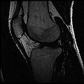
Ideal Spgr Fat MR image
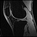
Ideal Spgr Water MR image
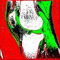
Segmentation Output


Key Investigators
- Stanford: Harish Doddi, Saikat Pal, Scott Delp
- Kitware: Luis Ibanez
Objective
The aim of this project is to develop an automatic/semi-automatic methodology to convert whole body imaging datasets into three-dimensional models for neuromuscular biomechanics and finite element simulations.
Approach, Plan
Our earlier work focused on segmenting the left atrium (LA), the heart chamber on which RF ablations are usually done, in blood pool MR images. We used a label fusion segmentation algorithm which first registered all of the training images to the test one and then employed a weighted voting procedure at each voxel.
Given corresponding cardiac blood pool and post-procedure delayed enhancement images for each patient, our plan is to first segment the LA in the blood pool image, then transfer this segmentation to the delayed enhancement image of the same patient. We intend to use this prior information while searching for the ablation scar using intensity based algorithms. This prior knowledge of the LA location will allow us to avoid most false positives.
Progress
We have only done some very preliminary ablation scar segmentation experiments.