Difference between revisions of "2010 Winter Project Week Musco Skeletal Segmentation"
From NAMIC Wiki
| Line 30: | Line 30: | ||
<h3>Approach, Plan</h3> | <h3>Approach, Plan</h3> | ||
| − | |||
| − | |||
</div> | </div> | ||
| Line 39: | Line 37: | ||
<h3>Progress</h3> | <h3>Progress</h3> | ||
| − | + | ||
</div> | </div> | ||
</div> | </div> | ||
Revision as of 22:32, 17 December 2009
Home < 2010 Winter Project Week Musco Skeletal Segmentation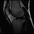
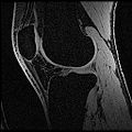
Ideal Spgr Fat MR image
Ideal Spgr Water MR image
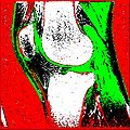
Segmentation Output
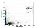
Scatter Plot
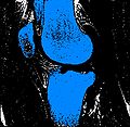
Bones
Cartilage

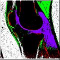
Slicer Output
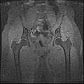
SpgrWater_Hip
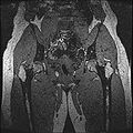
SpgrFat_Hip
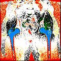
Hip Output

Hip Output


Key Investigators
- Stanford: Harish Doddi, Saikat Pal, Scott Delp
- Kitware: Luis Ibanez
Objective
The aim of this project is to develop an automatic/semi-automatic methodology to convert whole body imaging datasets into three-dimensional models for neuromuscular biomechanics and finite element simulations.
Approach, Plan
Progress