Difference between revisions of "2010 Winter Project Week Musco Skeletal Segmentation"
| Line 27: | Line 27: | ||
<h3>Approach, Plan</h3> | <h3>Approach, Plan</h3> | ||
| − | Approach: | + | <i><u>Approach:</u></i> |
<BR> | <BR> | ||
Multi-Contrast MR images are collected and seed points for each region of interest are taken as input. Cluster center and standard deviation are collected for each ROI based on pixel intensities of the seed points. The pixels are clustered based on different pixel intensity values in multiple MR images to the nearest cluster center radius. | Multi-Contrast MR images are collected and seed points for each region of interest are taken as input. Cluster center and standard deviation are collected for each ROI based on pixel intensities of the seed points. The pixels are clustered based on different pixel intensity values in multiple MR images to the nearest cluster center radius. | ||
<BR> | <BR> | ||
| − | Plan: | + | <i><u>Plan: </u></i> |
| + | <BR> | ||
a. Smooth the existing segmented label maps | a. Smooth the existing segmented label maps | ||
<BR> | <BR> | ||
Revision as of 20:38, 18 December 2009
Home < 2010 Winter Project Week Musco Skeletal Segmentation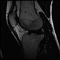
Ideal Spgr Fat MR image
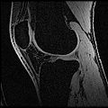
Ideal Spgr Water MR image
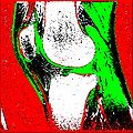
Segmentation Output
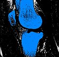
Bones
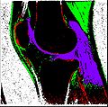
Cartilage
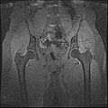
SpgrWater_Hip
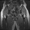
SpgrFat_Hip
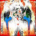
Hip Output


Key Investigators
- Stanford: Harish Doddi, Saikat Pal, Scott Delp
- Kitware: Luis Ibanez
Objective
The aim of this project is to develop an semi-automatic rapid methodology to convert whole body imaging datasets into three-dimensional geometries for modeling simulations. We have used a multi-contrast MR Segmentation approach to cluster the structures of interest. The output label maps will be smoothed and existing surface meshes will be morphed on to a target image.
Approach, Plan
Approach:
Multi-Contrast MR images are collected and seed points for each region of interest are taken as input. Cluster center and standard deviation are collected for each ROI based on pixel intensities of the seed points. The pixels are clustered based on different pixel intensity values in multiple MR images to the nearest cluster center radius.
Plan:
a. Smooth the existing segmented label maps
b. Build models for bones and cartilage from the existing segmented label maps.
Progress