Difference between revisions of "2010 Winter Project Week Spine Segmentation Module in Slicer3"
Sylvainjaume (talk | contribs) (New banner screenshots with text and arrows) |
|||
| Line 1: | Line 1: | ||
__NOTOC__ | __NOTOC__ | ||
<gallery> | <gallery> | ||
| − | Image:Loepprich_Jaume_vertebra_Slicer_Jan2010.png|Our | + | Image:Loepprich_Jaume_vertebra_Slicer_Jan2010.png|Our 3DSlicer module provides an automated segmentation of the spine in view of the treatment of disc herniation. [http://www.youtube.com/watch?v=kkAuP80kE8E (video)] |
| − | Image: | + | Image:Sylvain_Jaume_vertebrae_and_discs_Jan2010.png|This 3D rendering shows the nerve bundles surrounded by the cerebro-spinal fluid as they traverse the vertebrae. |
| − | Image: | + | Image:Sylvain_Jaume_disc_herniation_Jan2010.png|Back pain is caused by a pathological disc that protrudes into the vertebral foramen and pinches the nerve bundle exiting the spine. |
| − | Image:PW-SLC2010.png| | + | Image:PW-SLC2010.png|Our 3DSlicer module for automated segmentation has been presented at the [[2010_Winter_Project_Week#Projects|NA-MIC Project Week]] in Salt Lake City, Jan 4-8, 2010. |
</gallery> | </gallery> | ||
==Key Investigators== | ==Key Investigators== | ||
* Sylvain Jaume (MIT) | * Sylvain Jaume (MIT) | ||
| − | * Martin | + | * Martin Löpprich (University of Heidelberg) |
* Ron Kikinis, Steve Pieper (BWH) | * Ron Kikinis, Steve Pieper (BWH) | ||
* Polina Golland (MIT), Ehud Schmidt (BWH) | * Polina Golland (MIT), Ehud Schmidt (BWH) | ||
| Line 19: | Line 19: | ||
We are developing a Slicer module to segment the region within the thecal sac in MRI images of the spine. | We are developing a Slicer module to segment the region within the thecal sac in MRI images of the spine. | ||
Our objective is to provide a segmentation and visualization tool to improve the treatment of disc herniation. | Our objective is to provide a segmentation and visualization tool to improve the treatment of disc herniation. | ||
| − | The structures of interests are the cerebro-spinal fluid (CSF), the discs, the vertebrae and the spinal nerves | + | The structures of interests are the cerebro-spinal fluid (CSF), the discs, the vertebrae and the spinal nerves. |
</div> | </div> | ||
| Line 42: | Line 42: | ||
<div style="width: 97%; float: left;"> | <div style="width: 97%; float: left;"> | ||
| − | === | + | ===Illustrations=== |
| + | |||
| + | The cause of back pain is the pressure of the herniated disc onto the spinal nerves. The herniated disc pushes the canal for the cerebro-spinal fluid (CSF). The resulting pressure on the nerve bundles can cause severe discomfort and even permanent disability if not treated. | ||
{| | {| | ||
| − | |[[Image:spine_segmentation_module_in_slicer_001.png|thumb|300px| | + | |[[Image:spine_segmentation_module_in_slicer_001.png|thumb|300px|The segmentation the spine overlaid on the MRI image can help the surgeon during a minimally invasive intervention on the herniated disc.]] |
| − | |[[Image:spine_segmentation_module_in_slicer_002.png|thumb|300px| | + | |[[Image:spine_segmentation_module_in_slicer_002.png|thumb|300px|This Slicer rendering shows an herniated disc that protrudes into the vertebral foramen and reduces the opening for the nerve bundles and the cerebro-spinal fluid (CSF).]] |
|} | |} | ||
| + | |||
| + | The CSF has a double role: it acts as a lubricant between the vertebrae and as a mechanical insulation to protect the nerves from outside shocks. The main challenge is to perform the segmentation in a fully automated way. | ||
{| | {| | ||
Revision as of 08:10, 26 January 2010
Home < 2010 Winter Project Week Spine Segmentation Module in Slicer3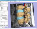
Our 3DSlicer module provides an automated segmentation of the spine in view of the treatment of disc herniation. (video)
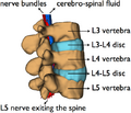
This 3D rendering shows the nerve bundles surrounded by the cerebro-spinal fluid as they traverse the vertebrae.
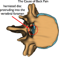
Back pain is caused by a pathological disc that protrudes into the vertebral foramen and pinches the nerve bundle exiting the spine.
Our 3DSlicer module for automated segmentation has been presented at the NA-MIC Project Week in Salt Lake City, Jan 4-8, 2010.


Key Investigators
- Sylvain Jaume (MIT)
- Martin Löpprich (University of Heidelberg)
- Ron Kikinis, Steve Pieper (BWH)
- Polina Golland (MIT), Ehud Schmidt (BWH)
Objective
We are developing a Slicer module to segment the region within the thecal sac in MRI images of the spine. Our objective is to provide a segmentation and visualization tool to improve the treatment of disc herniation. The structures of interests are the cerebro-spinal fluid (CSF), the discs, the vertebrae and the spinal nerves.
Approach, Plan
Our plan for the project week is to integrate our code into Slicer 3.5. Our code analyzes the intensity profile of different regions in the MRI and automatically defines the optimum region for the CSF.
Progress
The algorithm has been implemented in Slicer 3.5 as an extension module. This volumetric pattern recognition algorithm is fully implemented as a Slicer module using ITK and VTK. The module has been tested on data sets acquired at Brigham and Women's Hospital.
Illustrations
The cause of back pain is the pressure of the herniated disc onto the spinal nerves. The herniated disc pushes the canal for the cerebro-spinal fluid (CSF). The resulting pressure on the nerve bundles can cause severe discomfort and even permanent disability if not treated.


The CSF has a double role: it acts as a lubricant between the vertebrae and as a mechanical insulation to protect the nerves from outside shocks. The main challenge is to perform the segmentation in a fully automated way.


References
- Segmentation using Slicer 3.5, EMSegment module