Difference between revisions of "2011 Summer Project Week Contouring Gyne Structures"
| Line 29: | Line 29: | ||
<h3>Progress</h3> | <h3>Progress</h3> | ||
| − | + | *Used Grow-Cut Segmentation in Slicer. | |
| + | *[[Media:Gyn_brachy_grow_cut_segmentation.mov|Slicer Grow Cut Segmentation Movie]] | ||
Revision as of 01:25, 24 June 2011
Home < 2011 Summer Project Week Contouring Gyne Structures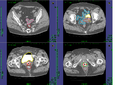
Anatomy Structures for Gynecological Brachytherapy


Project Title: Contouring of Anatomy Structures like Tumor, Bladder, Rectum and Sigmoid for Gynecological Brachytherapy
Key Investigators
- BWH: Tina Kapur, Akila Viswanathan
- University Hospital of Marburg: Jan Egger
Objective
This project aims to support the time consuming manual slice by slice segmentation process of contouring the anatomy structures for Gynecological Brachytherapy. Therefore (semi-)automated segmentation tools under the medical platform 3D Slicer are used to extract the anatomy structures like tumor, bladder, rectum and sigmoid. Furthermore, we want to evaluate the segmentation results achieved with the tools from 3D Slicer by using the manual segmentations.
Approach, Plan
For evaluation, we expect that we will be able to use the contours extracted by physicians with the Nucletron Oncentra Planning System.
References
- Gynecologic Radiation Therapy: Novel Approaches to Image-Guidance and Management. Akila N. Viswanathan, Christian Kirisits, Beth E. Erickson, and Richard Pötter. Springer. ISBN-13: 978-3540689546
Delivery Mechanism
The results will be provided as a technical report.