Difference between revisions of "2011 Winter Project Week:Intra-ProceduralProstateMotion"
| Line 5: | Line 5: | ||
Image:Transrectal_prostate_biopsy.jpg|Patient configuration for transrectal prostate biopsy in closed bore MRI | Image:Transrectal_prostate_biopsy.jpg|Patient configuration for transrectal prostate biopsy in closed bore MRI | ||
Image:Pelvis_and_prostate_motion_example.gif|Motion of the pelvis and prostate gland over the first 30 minutes of MR-guided transperineal biopsy procedure. | Image:Pelvis_and_prostate_motion_example.gif|Motion of the pelvis and prostate gland over the first 30 minutes of MR-guided transperineal biopsy procedure. | ||
| + | Image:TransrectalBiopsySagittal.png|Sagittal image taken during a transrectal MR-guided transrectal biopsy procedure. | ||
</gallery> | </gallery> | ||
Revision as of 18:28, 10 January 2011
Home < 2011 Winter Project Week:Intra-ProceduralProstateMotion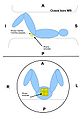
Patient configuration for transperineal prostate biopsy in closed bore MRI
Patient configuration for transrectal prostate biopsy in closed bore MRI
Motion of the pelvis and prostate gland over the first 30 minutes of MR-guided transperineal biopsy procedure.
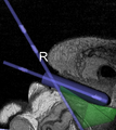
Sagittal image taken during a transrectal MR-guided transrectal biopsy procedure.


Key Investigators
- BWH: Andrey Fedorov
- Queens University: Andras Lasso
Objective
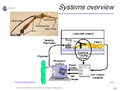
Multi-parametric diagnostic MRI was shown to be important in localization of prostate cancer. MR-guided prostate cancer biopsy (using transperineal access at BWH, transrectal access at Queen's clinical partners) performed in the closed bore MR scanner relies on image registration between the intra-procedural configuration of the prostate gland and the pre-procedural diagnostic MRI to locate the suspected cancer regions and guide the biopsy sample collection. Intra-operative image acquisition includes a volumetric scan of the whole prostate gland in the beginning of the procedure, followed by single-slice (or few orthogonal slices) acquisitions throughout the procedure with the purpose of confirming biopsy needle location. In the current image processing workflow, the diagnostic MRI is registered with the initial intra-procedural MRI.
One of the challenges we are facing is the motion and deformation of the prostate gland during the course of the procedure, which is typically ~1 hour long. Significant patient motion may invalidate the result of the initial pre- to intra-procedural MRI registration.
Our objective is to develop robust image acquisition and registration protocols to detect and compensate for the intra-procedural prostate motion/deformation, preferably using single-slice acquisitions throughout the procedure.
Approach, Plan
- evaluate and compare the solutions developed at BWH and Queens for volume-to-slice registration on the common data
- seek input from NA-MIC registration experts on the design of the registration approach
- discuss the high-level approach and plans for integrating volume-to-slice registration functionality into ProstateNav Slicer module
- discuss with Slicer engineering core plans and challenges in improving support for deformable transformations in 3D Slcer
Dissemination
http://viewvc.slicer.org/viewcvs.cgi/trunk/SLC2011-ProstateRegistration/?root=NAMICSandBox
References
- H. Tadayyon, A. Lasso, S. Gill, A. Kaushal, P. Guion, and G. Fichtinger, Target Motion Compensation in MRI-guided Prostate Biopsy with Static Images, EMBC2010 - 32nd Annual International Conference of the IEEE Engineering in Medicine and Biology Society, 2010
- H. Tadayyon, MRI-Guided Prostate Motion Tracking using Multislice-to-Volume Registration, MASc Thesis, 2010