Difference between revisions of "2011 Winter Project Week:RegistrationInPresenceOfAnatomicVariation"
(Created page with '__NOTOC__ <gallery> Image:PW-SLC2011.png|Projects List </gallery> ==Key Investigators== * Kitware: Danielle Pace, Stephen Aylward * UNC: M…') |
|||
| (7 intermediate revisions by one other user not shown) | |||
| Line 2: | Line 2: | ||
<gallery> | <gallery> | ||
Image:PW-SLC2011.png|[[2011_Winter_Project_Week#Projects|Projects List]] | Image:PW-SLC2011.png|[[2011_Winter_Project_Week#Projects|Projects List]] | ||
| + | Image:SlidingOrganRegistration_artificial.png|Example sliding organ registration on artificial images. Compared to the fixed image, the moving image translates the subset of the upper tube with increasing intensity to the right by four pixels, and the subset of the bottom tube with decreasing intensity to the left by four pixels. | ||
| + | Image:SlidingOrganRegistration_XCATimages.png|Models segmented from moving and fixed images created using the XCAT software phantom. | ||
| + | Image:SlidingOrganRegistration_XCATresults.png|Results of registering the XCAT phantom images: the anisotropic diffusive regularization produces a more physiologically realistic deformation field. | ||
</gallery> | </gallery> | ||
| Line 14: | Line 17: | ||
<h3>Objective</h3> | <h3>Objective</h3> | ||
| − | + | We are interested in registering images that contain anatomical variation between them, for example where organs slide against each other. For example, the brain may slide against the skull due to intraoperative brain shift, the heart slides relative to the lungs throughout the cardiac cycle, and respiration induces sliding of the lungs against the chest wall or abdominal muscles against each other. Not considering anatomical variation during image registration leads to errors that may impact clinical outcomes. | |
| + | |||
| + | We have developed a regularization approach for deformable image registration that considers sliding motion and is based on anisotropic diffusion, and have shown using artificial and phantom images that it results in more plausible deformation fields compared to a typical diffusive (Gaussian smoothing) regularization. | ||
</div> | </div> | ||
| Line 21: | Line 26: | ||
<h3>Approach, Plan</h3> | <h3>Approach, Plan</h3> | ||
| − | + | During the project week, we will discuss and prototype potential improvements to the method, which may include: | |
| + | * Organ border specification using the structure tensor (segmentation currently required) | ||
| + | * Incorporate more efficient diffusion calculation schemes / code profiling and optimization | ||
| + | * Representing normals as tensors rather than vectors, for improved performance at object corners | ||
</div> | </div> | ||
| Line 30: | Line 38: | ||
</div> | </div> | ||
| + | Very fruitful discussions on future work with Sandy Wells and Petter Risholm | ||
| + | |||
| + | Discussions and initial testing with Andriy Fedorov on applications to prostate interventions | ||
| + | |||
| + | Sliding organ registration profiling and optimizations: | ||
| + | |||
| + | - 18% faster in Debug mode | ||
| + | |||
| + | - 31% faster in Release mode | ||
| + | |||
| + | - (measured by registering two small 30x30x30 test images over 500 iterations) | ||
</div> | </div> | ||
| Line 35: | Line 54: | ||
==Delivery Mechanism== | ==Delivery Mechanism== | ||
| − | |||
| − | |||
| − | |||
| + | This work will be delivered to the NA-MIC Kit as a: | ||
| + | #ITK Module | ||
| + | #Slicer Module | ||
| + | ##Built-in | ||
| + | ##Extension -- commandline YES | ||
| + | ##Extension -- loadable | ||
| + | #Other YES | ||
| + | The sliding registration software is distributed as part of [http://public.kitware.com/Wiki/TubeTK TubeTK], a new open-source toolkit providing software for registration, segmentation, analysis and quantification of images depicting tubular stuctures, such as vessels, bronchi and neurons. In particular, the sliding organ registration algorithm is incorporated into 3D Slicer as a CLI ("Anisotropic Diffusive Deformable Registration") that is provided with TubeTK ([http://public.kitware.com/Wiki/TubeTK/Sliding_Organ_Registration see the algorithm's page here]). | ||
</div> | </div> | ||
Latest revision as of 17:53, 14 January 2011
Home < 2011 Winter Project Week:RegistrationInPresenceOfAnatomicVariation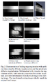
Example sliding organ registration on artificial images. Compared to the fixed image, the moving image translates the subset of the upper tube with increasing intensity to the right by four pixels, and the subset of the bottom tube with decreasing intensity to the left by four pixels.
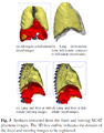
Models segmented from moving and fixed images created using the XCAT software phantom.
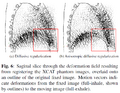
Results of registering the XCAT phantom images: the anisotropic diffusive regularization produces a more physiologically realistic deformation field.


Key Investigators
- Kitware: Danielle Pace, Stephen Aylward
- UNC: Marc Niethammer
- SPL: Sandy Wells, Tina Kapur, Petter Risholm
Objective
We are interested in registering images that contain anatomical variation between them, for example where organs slide against each other. For example, the brain may slide against the skull due to intraoperative brain shift, the heart slides relative to the lungs throughout the cardiac cycle, and respiration induces sliding of the lungs against the chest wall or abdominal muscles against each other. Not considering anatomical variation during image registration leads to errors that may impact clinical outcomes.
We have developed a regularization approach for deformable image registration that considers sliding motion and is based on anisotropic diffusion, and have shown using artificial and phantom images that it results in more plausible deformation fields compared to a typical diffusive (Gaussian smoothing) regularization.
Approach, Plan
During the project week, we will discuss and prototype potential improvements to the method, which may include:
- Organ border specification using the structure tensor (segmentation currently required)
- Incorporate more efficient diffusion calculation schemes / code profiling and optimization
- Representing normals as tensors rather than vectors, for improved performance at object corners
Progress
Very fruitful discussions on future work with Sandy Wells and Petter Risholm
Discussions and initial testing with Andriy Fedorov on applications to prostate interventions
Sliding organ registration profiling and optimizations:
- 18% faster in Debug mode
- 31% faster in Release mode
- (measured by registering two small 30x30x30 test images over 500 iterations)
Delivery Mechanism
This work will be delivered to the NA-MIC Kit as a:
- ITK Module
- Slicer Module
- Built-in
- Extension -- commandline YES
- Extension -- loadable
- Other YES
The sliding registration software is distributed as part of TubeTK, a new open-source toolkit providing software for registration, segmentation, analysis and quantification of images depicting tubular stuctures, such as vessels, bronchi and neurons. In particular, the sliding organ registration algorithm is incorporated into 3D Slicer as a CLI ("Anisotropic Diffusive Deformable Registration") that is provided with TubeTK (see the algorithm's page here).