Difference between revisions of "2011 Winter Project Week:The Vascular Modeling Toolkit in 3D Slicer"
(Created page with '__NOTOC__ <gallery> Image:3D_Segmentation_LA.png | 3D View of the Segmentation of Endocardial Wall Image:2d_axial_LA.png | 2D View </gallery> ==Key Investigators== * Georgia Tec…') |
|||
| (14 intermediate revisions by 3 users not shown) | |||
| Line 1: | Line 1: | ||
__NOTOC__ | __NOTOC__ | ||
<gallery> | <gallery> | ||
| − | Image: | + | Image:PW-SLC2011.png|[[2011_Winter_Project_Week#Projects|Projects List]] |
| − | Image: | + | Image:Slicervmtk logo.png|VMTK in 3D Slicer |
| + | Image:Vesselenhancement.png|Enhancing vasculature structures | ||
| + | Image:Easylevelsetsegmentation.png|Level-Set segmentation | ||
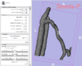
| + | Image:Centerlines.png|Computation of Centerlines | ||
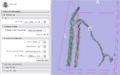
| + | Image:Networkextraction.png|Network extraction of vessel trees | ||
| + | Image:Branchsplitting.png|Branchsplitting | ||
| + | Image:Anim.gif|The new parameter widget for VMTKVesselness4 | ||
</gallery> | </gallery> | ||
==Key Investigators== | ==Key Investigators== | ||
| − | * | + | * UPenn: Daniel Haehn, Kilian Pohl |
| + | * Rutgers: M. Gokhan Uzunbas | ||
| + | * [http://www.orobix.com Orobix], Italy: Luca Antiga | ||
| + | * SPL: Steve Pieper, Ron Kikinis | ||
<div style="margin: 20px;"> | <div style="margin: 20px;"> | ||
| Line 12: | Line 21: | ||
<h3>Objective</h3> | <h3>Objective</h3> | ||
| + | The Vascular Modeling Toolkit ([http://www.vmtk.org VMTK]) is a collection of libraries and tools for 3D reconstruction, geometric analysis, mesh generation and surface data analysis for image-based modeling of blood vessels. | ||
| − | + | Several 3D Slicer extensions already exist and provide VMTK functionality in Slicer3. | |
| − | |||
| − | |||
| − | |||
</div> | </div> | ||
| Line 23: | Line 30: | ||
<h3>Approach, Plan</h3> | <h3>Approach, Plan</h3> | ||
| − | + | We want to investigate how to include the VMTK in 3D Slicer functionality in the upcoming Slicer4 application. This should include several enhancements to the existing user interfaces. | |
| − | We | ||
| − | |||
</div> | </div> | ||
| Line 31: | Line 36: | ||
<h3>Progress</h3> | <h3>Progress</h3> | ||
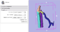
| + | '''Project Week Update:''' | ||
| + | Unfortunately, we did not have as much time as we liked to spend on this project. Nevertheless, we created a QT widget to optimize the selection of user-defined parameters for vessel enhancement. The new widget will be part of the upcoming first VMTK module for Slicer4 called VMTKVesselness4. | ||
| + | We plan to investigate if the integration of the new widget in CTK makes sense. | ||
</div> | </div> | ||
| Line 38: | Line 46: | ||
<div style="width: 97%; float: left;"> | <div style="width: 97%; float: left;"> | ||
| + | ==Delivery Mechanism== | ||
| + | |||
| + | This work will be delivered to the NA-MIC Kit as a (please select the appropriate options by noting YES against them below) | ||
| + | |||
| + | #ITK Module | ||
| + | #Slicer Module | ||
| + | ##Built-in | ||
| + | ##Extension -- commandline | ||
| + | ##Extension -- loadable [X] | ||
| + | #Other (Please specify) | ||
==References== | ==References== | ||
| − | * | + | * Antiga L, Piccinelli M, Botti L, Ene-Iordache B, Remuzzi A and Steinman DA. An image-based modeling framework for patient-specific computational hemodynamics. Medical and Biological Engineering and Computing, 46: 1097-1112, Nov 2008. |
| + | * D. Hähn. Integration of the vascular modeling toolkit in 3d slicer. SPL, 04 2009. Available online at http://www.spl.harvard.edu/publications/item/view/1728. | ||
| + | * D. Hähn. Centerline Extraction of Coronary Arteries in 3D Slicer using VMTK based Tools. Master's Thesis. Department of Medical Informatics, University of Heidelberg, Germany. Feb 2010. | ||
| + | * Piccinelli M, Veneziani A, Steinman DA, Remuzzi A, Antiga L (2009) A framework for geometric analysis of vascular structures: applications to cerebral aneurysms. IEEE Trans Med Imaging. In press. | ||
</div> | </div> | ||
Latest revision as of 15:37, 14 January 2011
Home < 2011 Winter Project Week:The Vascular Modeling Toolkit in 3D Slicer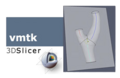
VMTK in 3D Slicer
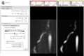
Enhancing vasculature structures
Level-Set segmentation
Computation of Centerlines

Network extraction of vessel trees
Branchsplitting
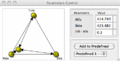
The new parameter widget for VMTKVesselness4


Key Investigators
- UPenn: Daniel Haehn, Kilian Pohl
- Rutgers: M. Gokhan Uzunbas
- Orobix, Italy: Luca Antiga
- SPL: Steve Pieper, Ron Kikinis
Objective
The Vascular Modeling Toolkit (VMTK) is a collection of libraries and tools for 3D reconstruction, geometric analysis, mesh generation and surface data analysis for image-based modeling of blood vessels.
Several 3D Slicer extensions already exist and provide VMTK functionality in Slicer3.
Approach, Plan
We want to investigate how to include the VMTK in 3D Slicer functionality in the upcoming Slicer4 application. This should include several enhancements to the existing user interfaces.
Progress
Project Week Update: Unfortunately, we did not have as much time as we liked to spend on this project. Nevertheless, we created a QT widget to optimize the selection of user-defined parameters for vessel enhancement. The new widget will be part of the upcoming first VMTK module for Slicer4 called VMTKVesselness4.
We plan to investigate if the integration of the new widget in CTK makes sense.
Delivery Mechanism
This work will be delivered to the NA-MIC Kit as a (please select the appropriate options by noting YES against them below)
- ITK Module
- Slicer Module
- Built-in
- Extension -- commandline
- Extension -- loadable [X]
- Other (Please specify)
References
- Antiga L, Piccinelli M, Botti L, Ene-Iordache B, Remuzzi A and Steinman DA. An image-based modeling framework for patient-specific computational hemodynamics. Medical and Biological Engineering and Computing, 46: 1097-1112, Nov 2008.
- D. Hähn. Integration of the vascular modeling toolkit in 3d slicer. SPL, 04 2009. Available online at http://www.spl.harvard.edu/publications/item/view/1728.
- D. Hähn. Centerline Extraction of Coronary Arteries in 3D Slicer using VMTK based Tools. Master's Thesis. Department of Medical Informatics, University of Heidelberg, Germany. Feb 2010.
- Piccinelli M, Veneziani A, Steinman DA, Remuzzi A, Antiga L (2009) A framework for geometric analysis of vascular structures: applications to cerebral aneurysms. IEEE Trans Med Imaging. In press.