Difference between revisions of "2011 Winter Project Week:TubeTK VascularImageSegmentationAndAnalysis"
From NAMIC Wiki
| (One intermediate revision by the same user not shown) | |||
| Line 1: | Line 1: | ||
__NOTOC__ | __NOTOC__ | ||
<gallery> | <gallery> | ||
| + | Image:TubeTK-VessTortuosity.jpg|Tortuosity for benign/malignant (Image from Dr. Bullitt, UNC) | ||
| + | Image:TubeTK-USVess.png|Ultrasound to vessel registration | ||
| + | Image:TubeTK-VessGraphs.jpg|Spatial graphs of vasculature capture inter-population variations | ||
Image:PW-SLC2011.png|[[2011_Winter_Project_Week#Projects|Projects List]] | Image:PW-SLC2011.png|[[2011_Winter_Project_Week#Projects|Projects List]] | ||
</gallery> | </gallery> | ||
| Line 18: | Line 21: | ||
* Surgical guidance: registering pre-operative vascular models with intra-operative images (e.g., ultrasound) | * Surgical guidance: registering pre-operative vascular models with intra-operative images (e.g., ultrasound) | ||
* Characterizing vascular patters: using graph theory to distinguish clinical populations based on vascular patterns (e.g., benign -vs- malignant tumors via tortuosity) | * Characterizing vascular patters: using graph theory to distinguish clinical populations based on vascular patterns (e.g., benign -vs- malignant tumors via tortuosity) | ||
| + | |||
| + | History | ||
| + | * June 2001, UNC released the patent on vessel extraction method from [Aylward, Bullitt 1996...] | ||
| + | * TubeTK released under Apache 2.0 license: includes rights to patents | ||
</div> | </div> | ||
| Line 33: | Line 40: | ||
<h3>Progress</h3> | <h3>Progress</h3> | ||
| + | * Skype meeting with VMTK team to learn design pattern to follow | ||
| + | * Extended TubeTK to include LDA methods for multi-echo MR segmentation | ||
| + | ** SWAN (susceptibility weighted angiography), T1, T2 data from U of Mississippi | ||
</div> | </div> | ||
Latest revision as of 17:32, 14 January 2011
Home < 2011 Winter Project Week:TubeTK VascularImageSegmentationAndAnalysis
Tortuosity for benign/malignant (Image from Dr. Bullitt, UNC)
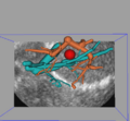
Ultrasound to vessel registration
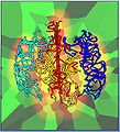
Spatial graphs of vasculature capture inter-population variations


Key Investigators
- Kitware: Stephen Aylward, Danielle Pace
- SPL: Steve Pieper
- Luca Antiga, Daniel Haehn
Objective
TubeTK is a new open-source toolkit that hosts algorithms for applications involving images of tubes.
Two driving applications:
- Surgical guidance: registering pre-operative vascular models with intra-operative images (e.g., ultrasound)
- Characterizing vascular patters: using graph theory to distinguish clinical populations based on vascular patterns (e.g., benign -vs- malignant tumors via tortuosity)
History
- June 2001, UNC released the patent on vessel extraction method from [Aylward, Bullitt 1996...]
- TubeTK released under Apache 2.0 license: includes rights to patents
Approach, Plan
- Python module in Slicer 4 for centerline and radius estimation of vasculature in brain MRA
- Workflow: brain envelop segmentation, seeding, extraction
- Integration with VMTK
Progress
- Skype meeting with VMTK team to learn design pattern to follow
- Extended TubeTK to include LDA methods for multi-echo MR segmentation
- SWAN (susceptibility weighted angiography), T1, T2 data from U of Mississippi
Delivery Mechanism
This work will be delivered to the NA-MIC Kit as follows:
- All software written during the project week will be contributed to TubeTK, and algorithms will be incorporated into 3D Slicer as CLI applications.