Difference between revisions of "2013 Summer Project Week:CARMA AutoLASeg"
From NAMIC Wiki
m |
|||
| Line 31: | Line 31: | ||
<h3>Progress</h3> | <h3>Progress</h3> | ||
| − | This module has been nearly completed, and we need to improve the visualization of the final isosurface of the left atrium. | + | This module has been nearly completed, and we need to improve the visualization of the final isosurface of the left atrium. We also need to find a way to store and access (through Slicer) a large amount of training model data. |
</div> | </div> | ||
Revision as of 03:00, 17 June 2013
Home < 2013 Summer Project Week:CARMA AutoLASegKey Investigators
- Utah: Salma Bengali, Alan Morris, Josh Cates, Gopal Veni, Ross Whitaker, Rob MacLeod
Objective
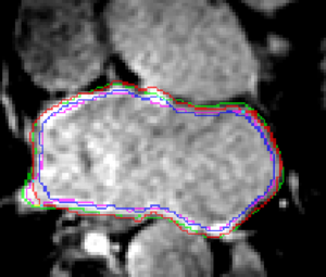
The goal of this project is to implement Gopal and Ross' segmentation method: "Left atrial wall segmentation using intensity profile based feature detector and optimal graph-cuts." Our objective is to implement this method in Slicer. There are several fairly involved steps for the entire segmentation process, including:
- An LA shape model building phase
- User input to identify the center of a region of interest
- Computation of the segmentation, given a new LGE-MRI image
- Surface reconstruction of the output point set and (optionally) scan-conversion of the surface mesh to a binary segmentation volume.
Approach, Plan
We plan to implement this algorithm as a Slicer command-line module.
Progress
This module has been nearly completed, and we need to improve the visualization of the final isosurface of the left atrium. We also need to find a way to store and access (through Slicer) a large amount of training model data.
Delivery Mechanism
This work will be delivered to the NA-MIC Kit as a Slicer module as part of the existing Cardiac MRI Toolkit extension.

