|
|
| Line 33: |
Line 33: |
| | | | |
| | = Research Plan = | | = Research Plan = |
| − | The overall goal of the project is to improve the capabilities for
| |
| − | *Goal: Scientific evidence for or against adaptive proton-beam radiotherapy in the base of skull region
| |
| − | *Require: Update literature review
| |
| − | *Recommend: Study to quantify geometry of anatomic differences
| |
| − | *Require: Comparison study of delivered vs. planned dose
| |
| − | *Require: Comparison study of delivered vs. adaptive dose
| |
| − | *Wishlist: Comparison study of IMRT vs. protons
| |
| − |
| |
| − | === Engineering ===
| |
| − | See: http://www.na-mic.org/Wiki/index.php/EngineeringRetreat2010
| |
| − |
| |
| − | *[[Projects:DBP3:MGH_CMake_Enhancements|Pushing CMake module bug fixes upstream]]
| |
| − | *Window/level slider bug [http://www.na-mic.org/Bug/view.php?id=1047 1047]
| |
| − | *Add mode compositing glitches [http://www.na-mic.org/Bug/view.php?id=1048 1048] [http://www.na-mic.org/Bug/view.php?id=1049 1049]
| |
| − |
| |
| − | === Image Registration ===
| |
| − | *Goal: Multiple slicer plugins for registration
| |
| − | *Recommend: Experimental evaluation of existing registration methods
| |
| − | *Require: Interactive registration (landmark splines)
| |
| − | *Recommend: Regularized B-splines
| |
| − | *Recommend: B-splines with landmark constraints
| |
| − | *Recommend: B-splines with surface constraints
| |
| − | *Wishlist: Parameter-free registration
| |
| − | *Wishlist: Image-free surface registration
| |
| − | *Wishlist: Parallel optimization
| |
| − |
| |
| − | === Image Segmentation ===
| |
| − | *Goal: Slicer plugin for automatic segmentation
| |
| − | *Require: Contour propagation for intra-subject segmentation
| |
| − | *Recommend: Automatic segmentation (atlas-based) for inter-subject segmentation
| |
| − | *Recommend: Automatic segmentation (model-based) for inter-subject segmentation
| |
| − | *Wishlist: Contour interpolation methods for interactive segmentation
| |
| − |
| |
| − | === Radiotherapy Workflow ===
| |
| − | *Goal: Reasonably complete workflow, including dose review and comparison tools
| |
| − | *Require: Labelmap that can handle overlapping structures
| |
| − | *Require: Visualization of RT dose (2D, as isodose lines, with legend)
| |
| − | *Wishlist: Visualization of RT dose (3D, w/ Shadie)
| |
| − | *Require: DICOM improvements (CT export, DICOM-RT import/export)
| |
| − | *Require: Compute dose volume histograms
| |
| − | *Recommend: Margin tools
| |
| − | *Wishlist: Visualize dose volume histograms
| |
| − | *Require: Better support for extensions
| |
| − | *Recommend: MRML node for registration
| |
| − | *Recommend: MRML node for patient demographics
| |
| − | *Wishlist: Unevely spaced CT
| |
| − | *Wishlist: Vector field visualization
| |
| − | *Wishlist: DICOM Network I/O
| |
| − |
| |
| − | === Data ===
| |
| − | * Goal: 50 cases for inter-subject segmentation
| |
| − | * Goal: 30 cases for intra-subject registration
| |
| − | * Require: New retrospective IRB
| |
| − | * Require: Careful QA of segmentation data
| |
| − | * Require: Validation data for registration studies
| |
| − |
| |
| − | === Outreach ===
| |
| − | *Require: Symposium or tutorial at major conference
| |
| − | *Require: Hands-on tutorial at MGH
| |
| − | *Require: On-line tutorials for radiotherapy workflow
| |
| − | *Recommend: User's group meeting at major conference
| |
| | | | |
| | = Key Personnel = | | = Key Personnel = |
Back to NA-MIC DBPs | NA-MIC Cores
Introduction
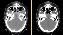
Anatomic change due to tumor regression in nasopharyngeal cancer
Head and neck cancers account for about 60,000 new cancer cases per year and represent about 6% of all cancers in the United States. These cancers are treated by a combination of chemotherapy, radiotherapy, and surgery. The five-year survival is approximately 50%. During a six-week regimen of radiotherapy, head and neck cancer patients often exhibit anatomic changes that affect their treatment. These changes include tumor regression or growth, changes in lymph node size, and changes in air cavities. Uncorrected, these changes can increase the risk of treatment complications or reduce treatment efficacy.
Adaptive radiotherapy is a form of personalized medicine that addresses the problem of anatomic change by incrementally adjusting the radiotherapy plan. However, a mid-treatment adjustment is complex: it requires a new CT image, image segmentation, deformable registration, and mapping of the previously delivered dose onto the new image. This project proposes to use the NA-MIC Kit to develop a simple, practical workflow for achieving adaptive radiotherapy which can be applied on a case-by-case basis.
For details on the background and goals, please see the project web page.
For a more extensive wishlist, see the planning meeting notes
Project Goals
This project has three specific aims:
- Develop an open computational workflow for adaptive radiotherapy.
- Validate the accuracy of image analysis algorithms for radiotherapy.
- Evaluate the dosimetric gain of adaptive radiotherapy.
External-beam radiotherapy requires a unique set of image analysis tools, which are only partly supported in the NA-MIC kit. Our specific aims require us to broaden the capabilities of 3D Slicer to support data interchange with commercial software, and to manipulate, review, and validate radiotherapy planning objects. We specify four sub-goals which we will work toward:
DICOM-RT Interchange
Structure and Dose Warping
Plan Review
Validation Tools
State of the Art: 2010
Research Plan
Key Personnel
- MGH: Greg Sharp, Annie Chan, George TY Chen, Nadya Shusharina, Rui Li, Ken Westover, Itai Pashtan, John Wolfgang
- MIT: Polina Golland, Michal Depa
- GT: Allen Tannenbaum, Ivan Kolesov
- Isomics: Steve Pieper
