Difference between revisions of "Projects:RegistrationLibrary:RegLib C05"
From NAMIC Wiki
m (Text replacement - "http://www.slicer.org/slicerWiki/index.php/" to "https://www.slicer.org/wiki/") |
|||
| (14 intermediate revisions by one other user not shown) | |||
| Line 3: | Line 3: | ||
[[Projects:RegistrationDocumentation:UseCaseInventory|Back to Registration Use-case Inventory]] <br> | [[Projects:RegistrationDocumentation:UseCaseInventory|Back to Registration Use-case Inventory]] <br> | ||
| − | |||
| − | {| style="color:#bbbbbb | + | = <small>v3.6.1</small> [[Image:Slicer3-6Announcement-v1.png|150px]] Slicer Registration Library Case 05: <br> Inter-subject Knee MRI = |
| − | |[[Image:RegLib C05 KneeMRI1.png| | + | === Input === |
| − | |[[Image: | + | {| style="color:#bbbbbb; " cellpadding="10" cellspacing="0" border="0" |
| − | |[[Image:RegLib C05 KneeMRI2.png| | + | |[[Image:RegLib C05 KneeMRI1.png|150px|lleft|this is the main fixed reference image. All images are ev. aligned into this space]] |
| − | + | |[[Image:RegArrow_Affine.png|70px|lleft]] | |
| − | + | |[[Image:RegLib C05 KneeMRI2.png|150px|lleft|this is the moving image]] | |
| − | |||
|- | |- | ||
| − | | | + | |subject 1 |
| | | | ||
| − | | | + | |subject 1 |
| − | |||
| − | |||
| − | |||
| − | |||
|} | |} | ||
| + | |||
| + | === Modules === | ||
| + | *'''Slicer 3.6.1 recommended modules: [https://www.slicer.org/wiki/Modules:PythonSurfaceICPRegistration-Documentation-3.6 Surface Registration] | ||
| + | |||
===Objective / Background === | ===Objective / Background === | ||
| − | The final goal is to align a segmentation prior model to aid in cartilage segmentation. | + | The final goal is to align a segmentation prior model/atlas to aid in cartilage segmentation. |
| + | |||
=== Keywords === | === Keywords === | ||
MRI, knee, inter-subject, segmentation | MRI, knee, inter-subject, segmentation | ||
===Input Data=== | ===Input Data=== | ||
| − | * | + | *fixed: T1 SPGR , 0.9375 x 0.9375 x 1.4 mm voxel size, axial. + surface model from femur & tibia segmentation. |
| − | * | + | *moving: T1 SPGR , 0.9375 x 0.9375 x 1.2 mm voxel size, sagittal. + surface model from femur & tibia segmentation. |
| − | + | === Procedure / Pipeline === | |
| − | + | #Open case scene file or import image data + surface models | |
| − | + | #Open ''Registration / Surface Registration'' module | |
| + | ##Input Surface: S02_femur | ||
| + | ##Target Surface: S01_femur | ||
| + | ##Output Transform: create new transform, rename to "Xf1_surf_S2-S1" | ||
| + | ##Select/check ''Mean distance mode: RMS'' | ||
| + | ##Leave defaults: iterations: 50, landmarks: 200 | ||
| + | ##Select/check ''Start by matching centroids'' | ||
| + | ##Select/check ''Check mean distance'' | ||
| + | ##Click: ''Apply''; process duration: ~ seconds | ||
| + | #Go to the ''Data'' module | ||
| + | ##find the newly created transform node "Xf1_surf_S2-S1" and drag the "S02_femur" model node (and all other S02 entities) inside the transform. | ||
| + | ##for the 3D viewer turn on the visibility of the surface models (should be on already) | ||
| + | ##the surfaces for the two femur models should now align in basic position and orientation | ||
| + | #for additional details see tutorial downloads below | ||
| + | |||
=== Registration Results=== | === Registration Results=== | ||
| − | + | [[Image:RegLib C05 AGif reg.gif|200px|femur surface models before/after registration]] | |
| − | | | ||
===Download === | ===Download === | ||
| − | * | + | *Data |
| − | + | **[[Media:RegLib_C05_Data.zip|'''RegLib_C05_Data''': Example Dataset <small> (MRI images, surface models,Presets, Solution, zip file 9.5 MB) </small>]] | |
| − | < | + | *Presets |
| − | + | **[[Media:RegLib_C05_Presets.mrml|'''RegLib_C05_Presets.mrml''': registration parameter presets<small> (.mrml text file 12 kB) </small>]] | |
| − | + | *[[Projects:RegistrationDocumentation:ParameterPresetsTutorial|Link to User Guide: How to Load/Save Registration Parameter Presets]] | |
| − | + | *Documentation | |
| − | + | **[[Media:RegLib_C05_Tutorial.ppt|'''RegLib_C05_Tutorial''': Powerpoint file <small> (.ppt file 1 MB) </small>]] | |
| − | + | **[[Media:RegLib_C05_Tutorial.ppt|'''RegLib_C05_Tutorial''': PDF file <small> (.pdf file 0.5 MB) </small>]] | |
| − | * | ||
| − | * | ||
| − | |||
| − | |||
| − | === | + | === Acknowledgments === |
| − | |||
| − | |||
| − | |||
| − | |||
| − | |||
| − | |||
| − | |||
| − | |||
Latest revision as of 17:06, 10 July 2017
Home < Projects:RegistrationLibrary:RegLib C05Back to ARRA main page
Back to Registration main page
Back to Registration Use-case Inventory
Contents
v3.6.1 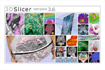 Slicer Registration Library Case 05:
Inter-subject Knee MRI

Input

|

|

|
| subject 1 | subject 1 |
Modules
- Slicer 3.6.1 recommended modules: Surface Registration
Objective / Background
The final goal is to align a segmentation prior model/atlas to aid in cartilage segmentation.
Keywords
MRI, knee, inter-subject, segmentation
Input Data
- fixed: T1 SPGR , 0.9375 x 0.9375 x 1.4 mm voxel size, axial. + surface model from femur & tibia segmentation.
- moving: T1 SPGR , 0.9375 x 0.9375 x 1.2 mm voxel size, sagittal. + surface model from femur & tibia segmentation.
Procedure / Pipeline
- Open case scene file or import image data + surface models
- Open Registration / Surface Registration module
- Input Surface: S02_femur
- Target Surface: S01_femur
- Output Transform: create new transform, rename to "Xf1_surf_S2-S1"
- Select/check Mean distance mode: RMS
- Leave defaults: iterations: 50, landmarks: 200
- Select/check Start by matching centroids
- Select/check Check mean distance
- Click: Apply; process duration: ~ seconds
- Go to the Data module
- find the newly created transform node "Xf1_surf_S2-S1" and drag the "S02_femur" model node (and all other S02 entities) inside the transform.
- for the 3D viewer turn on the visibility of the surface models (should be on already)
- the surfaces for the two femur models should now align in basic position and orientation
- for additional details see tutorial downloads below
Registration Results

Download
- Data
- Presets
- Link to User Guide: How to Load/Save Registration Parameter Presets
- Documentation