Difference between revisions of "Projects:RegistrationLibrary:RegLib C20"
From NAMIC Wiki
(Created page with 'Back to ARRA main page <br> Back to Registration main page <br> [[Projects:RegistrationDocumentation:UseCaseInv…') |
|||
| Line 45: | Line 45: | ||
**[[Projects:RegistrationDocumentation:ParameterPresetsTutorial|Link to User Guide: How to Load/Save Registration Parameter Presets]] | **[[Projects:RegistrationDocumentation:ParameterPresetsTutorial|Link to User Guide: How to Load/Save Registration Parameter Presets]] | ||
| − | === | + | === Procedures=== |
| − | *'''Phase | + | *'''Phase 1''': rigid alignment |
| − | #the | + | #Go to the [http://www.slicer.org/slicerWiki/index.php/Modules:BRAINSFit '''BRAINSfit'''] module |
| − | + | ##select Presets "Xf1_Rigid" or "Xf2_Affine" or set the parameters as given below: | |
| − | ## | + | ##fixed image: "CT_1", moving image: "CT_2" |
| − | ## | + | ##''Initialize with previous transform'': select "Off" |
| − | + | ##''Initialize Transform Mode'': check box for ''use MomentsAlign'' | |
| − | # | + | ##check ''Include Rigid registration Phase'' box. |
| − | ## | + | ##Output: under ''Slicer Linear Transform'', select new and rename to "Xf1_Rigid" or similar |
| − | ## | + | ##Registration Parameters: set "Number of Samples" to 200,000 at least |
| − | + | ##leave rest at defaults | |
| − | + | ##Click Apply | |
| − | *'''Phase | + | #return to the [http://www.slicer.org/slicerWiki/index.php/Modules:Data-Documentation-3.6 Data module] and drag the CT_2 inside/outside the different registration transforms to compare the alignment |
| − | # | + | *'''Phase 2''': affine alignment |
| − | ## | + | #Go to the [http://www.slicer.org/slicerWiki/index.php/Modules:BRAINSFit '''BRAINSfit'''] module |
| − | ## | + | ##same as Phase 1 above, except: |
| − | + | ##''Initialize with previous transform'': select "Xf1_Rigid" from phase 1 above | |
| − | ## | + | ##''Initialize Transform Mode'': check box for ''Off'' |
| − | + | ##Output: under ''Slicer Linear Transform'', select new and rename to "Xf2_Affine" or similar | |
| − | + | *'''Phase 3''': BSpline alignment | |
| − | + | ##same as Phase 2 above, except: | |
| − | + | ##''Initialize with previous transform'': select "Xf2_Affine" from phase 2 above | |
| − | + | ##Output: under ''Slicer BSpline Transform'', select new and rename to "Xf3_BSpline" or similar | |
| − | ## | + | ##Output Image Volume: select new and rename to "CT_2_Xf3" or similar |
| − | *'''Phase | + | ##Registration Parameters: set "Number of Samples" to 250,000 at least |
| − | + | ##Number of Grid Subdivisions: 9,9,5 or 11,11,7 | |
| − | ## | + | ##Click Apply |
| − | ## | + | #return to the [http://www.slicer.org/slicerWiki/index.php/Modules:Data-Documentation-3.6 Data module] and drag the CT_2 inside/outside the different registration transforms to compare the alignment |
| − | + | #to obtain a resampled volume: move CT_2 inside Xform of choice and then right-click on the volume and select ''Harden Transforms''. Save MRI under new name. | |
| − | ##Output | + | *'''Phase 4''': Resample PET |
| − | ##Output Volume: | + | #Go to the [http://www.slicer.org/slicerWiki/index.php/Modules:ResampleScalarVectorDWIVolume-Documentation-3.6 '''ResampleScalarVectorDWIVolume'''] module |
| − | ## | + | ##''Input Volume'': PET_2 ; ''Reference Volume'': PET_1 ; ''Output Volume'': create new, rename to "PET_2_Xf3" |
| − | ## | + | ##''Transform Node'': select "Xf3_BSpline" created in phase 3 above |
| − | + | ##''Transform Order'': check box for "output-to-input" | |
| − | *'''Phase | + | ##Click Apply |
| − | #Go to '' | ||
| − | ## | ||
| − | |||
| − | |||
| − | |||
| − | |||
| − | ## | ||
| − | ## | ||
| − | |||
| − | ##Click | ||
| − | |||
| − | |||
<!-- | <!-- | ||
| Line 99: | Line 87: | ||
=== Discussion: Registration Challenges === | === Discussion: Registration Challenges === | ||
*accuracy is the critical criterion here. We need the registration error (residual misalignment) to be smaller than the change we want to measure/detect. Agreement on what constitutes good alignment can therefore vary greatly. | *accuracy is the critical criterion here. We need the registration error (residual misalignment) to be smaller than the change we want to measure/detect. Agreement on what constitutes good alignment can therefore vary greatly. | ||
| − | |||
*because of the large FOV we have strong non-rigid deformations from differences in patient/limb positions etc. | *because of the large FOV we have strong non-rigid deformations from differences in patient/limb positions etc. | ||
*images are large volumes (>100 MB total) | *images are large volumes (>100 MB total) | ||
| − | |||
*2 images pairs have to be aligned, i.e. the calculated transform must be applied to the second (PET) image. | *2 images pairs have to be aligned, i.e. the calculated transform must be applied to the second (PET) image. | ||
=== Discussion: Key Strategies === | === Discussion: Key Strategies === | ||
*to calculate the transform, we use the images with the most accurate geometric representation and the smallest expected change, i.e. we align the follow-up CT to the baseline CT and then apply the transforms to the PET image. | *to calculate the transform, we use the images with the most accurate geometric representation and the smallest expected change, i.e. we align the follow-up CT to the baseline CT and then apply the transforms to the PET image. | ||
*because of the non-rigid differences due to posture and breathing we will need to apply a 2-step registration with an affine alignment followed by a BSpline. | *because of the non-rigid differences due to posture and breathing we will need to apply a 2-step registration with an affine alignment followed by a BSpline. | ||
| − | |||
| − | |||
| − | |||
| − | |||
Revision as of 18:33, 5 September 2011
Home < Projects:RegistrationLibrary:RegLib C20Back to ARRA main page
Back to Registration main page
Back to Registration Use-case Inventory
Contents
v3.6.3  Slicer Registration Library Case #20: Intra-subject whole-body PET-CT

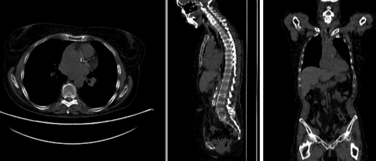
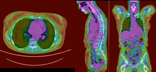
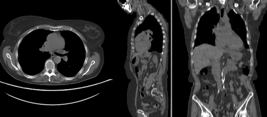
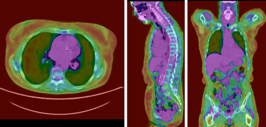
Input

|

|

|
| fixed image/target | moving image |
Modules
- Slicer 3.6.3 recommended modules: BrainsFit,
Objective / Background
Change assessment.
Keywords
PET-CT, whole-body, change assessment
Input Data
- reference/fixed : baseline CT: 0.98 x 0.98 x 5 mm , 512 x 512 x 149; PET: 4.1 x 4.1 x 5 mm , 168 x 168 x 149
- moving: CT: 0.98 x 0.98 x 5 mm , 512 x 512 x 149; PET: 4.1 x 4.1 x 5 mm , 168 x 168 x 149
Registration Results
rigid
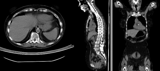
affine alignment
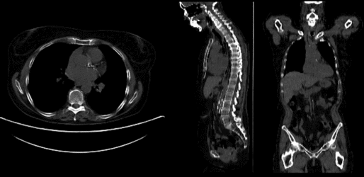
BSpline registration of full volumes. 9 x 9 x 5 grid
PET overlay ; BSpline registration of full volumes. 9 x 9 x 5 grid
BSpline registration of full volumes. 11 x 11 x 7 grid
PET overlay ; BSpline registration of full volumes. 11 x 11 x 7 grid


Download
- Data
Procedures
- Phase 1: rigid alignment
- Go to the BRAINSfit module
- select Presets "Xf1_Rigid" or "Xf2_Affine" or set the parameters as given below:
- fixed image: "CT_1", moving image: "CT_2"
- Initialize with previous transform: select "Off"
- Initialize Transform Mode: check box for use MomentsAlign
- check Include Rigid registration Phase box.
- Output: under Slicer Linear Transform, select new and rename to "Xf1_Rigid" or similar
- Registration Parameters: set "Number of Samples" to 200,000 at least
- leave rest at defaults
- Click Apply
- return to the Data module and drag the CT_2 inside/outside the different registration transforms to compare the alignment
- Phase 2: affine alignment
- Go to the BRAINSfit module
- same as Phase 1 above, except:
- Initialize with previous transform: select "Xf1_Rigid" from phase 1 above
- Initialize Transform Mode: check box for Off
- Output: under Slicer Linear Transform, select new and rename to "Xf2_Affine" or similar
- Phase 3: BSpline alignment
- same as Phase 2 above, except:
- Initialize with previous transform: select "Xf2_Affine" from phase 2 above
- Output: under Slicer BSpline Transform, select new and rename to "Xf3_BSpline" or similar
- Output Image Volume: select new and rename to "CT_2_Xf3" or similar
- Registration Parameters: set "Number of Samples" to 250,000 at least
- Number of Grid Subdivisions: 9,9,5 or 11,11,7
- Click Apply
- return to the Data module and drag the CT_2 inside/outside the different registration transforms to compare the alignment
- to obtain a resampled volume: move CT_2 inside Xform of choice and then right-click on the volume and select Harden Transforms. Save MRI under new name.
- Phase 4: Resample PET
- Go to the ResampleScalarVectorDWIVolume module
- Input Volume: PET_2 ; Reference Volume: PET_1 ; Output Volume: create new, rename to "PET_2_Xf3"
- Transform Node: select "Xf3_BSpline" created in phase 3 above
- Transform Order: check box for "output-to-input"
- Click Apply
Discussion: Registration Challenges
- accuracy is the critical criterion here. We need the registration error (residual misalignment) to be smaller than the change we want to measure/detect. Agreement on what constitutes good alignment can therefore vary greatly.
- because of the large FOV we have strong non-rigid deformations from differences in patient/limb positions etc.
- images are large volumes (>100 MB total)
- 2 images pairs have to be aligned, i.e. the calculated transform must be applied to the second (PET) image.
Discussion: Key Strategies
- to calculate the transform, we use the images with the most accurate geometric representation and the smallest expected change, i.e. we align the follow-up CT to the baseline CT and then apply the transforms to the PET image.
- because of the non-rigid differences due to posture and breathing we will need to apply a 2-step registration with an affine alignment followed by a BSpline.