Difference between revisions of "Rhesus Juvenile WMGM Atlas"
From NAMIC Wiki
| Line 6: | Line 6: | ||
* T1 subject images acquired | * T1 subject images acquired | ||
| − | + | * The atlas construction procedure was adapted from Automatic brain segmentation in rhesus monkeys. Proc SPIE Medical Imaging Conference, M. Styner, R. Knickmeyer, S. Joshi, C. Coe, S. J. Short, and J. Gilmore. Proc SPIE Vol 6512 Medical Imaging 2007, pp 65122L-1 - 65122L-8. | |
<gallery caption="Juvenile Rhesus Probabilistic Atlas" widths="350px" heights="180px" perrow="2"> | <gallery caption="Juvenile Rhesus Probabilistic Atlas" widths="350px" heights="180px" perrow="2"> | ||
Revision as of 03:52, 13 February 2008
Home < Rhesus Juvenile WMGM AtlasObjective:
- Develop a juvenile WM/GM/CSF atlas from normal subject images.
Progress:
- T1 subject images acquired
- The atlas construction procedure was adapted from Automatic brain segmentation in rhesus monkeys. Proc SPIE Medical Imaging Conference, M. Styner, R. Knickmeyer, S. Joshi, C. Coe, S. J. Short, and J. Gilmore. Proc SPIE Vol 6512 Medical Imaging 2007, pp 65122L-1 - 65122L-8.
- Juvenile Rhesus Probabilistic Atlas
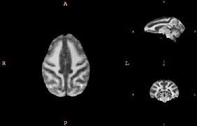
left
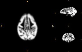
left
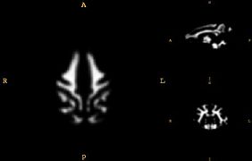
Probabilistic map of WM for Rhesus population


Key Investigators:
- Virginia Tech: Chris Wyatt and Jenny Long
Acknowledgement
- Linda Porrino, Wake Forest University