Difference between revisions of "User:Noby"
| Line 40: | Line 40: | ||
# Incorporating fMRI data using image registration and thresholding | # Incorporating fMRI data using image registration and thresholding | ||
# Creating a 3D model of the tumour volume | # Creating a 3D model of the tumour volume | ||
| − | * 3:30 - | + | *3:30 - 3:45 pm Coffee-break |
| − | *Module 4-6 ( | + | *Module 4-6 (3:45-4:45 pm) (TBD) |
# Predicting the locations of brain structures using image registration and a brain atlas | # Predicting the locations of brain structures using image registration and a brain atlas | ||
# Incorporating brain fiber tractography from diffusion weighted images | # Incorporating brain fiber tractography from diffusion weighted images | ||
Revision as of 03:01, 9 February 2010
M==Course Title==
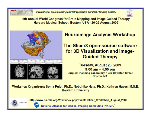
Introduction to Opensource Software Slicer and its Application to Image-Guided Therapy
Contents
Course Syllabus
The purpose of this tutorial is to provide the members of the research community with a practical experience of the image processing, 3D visualization, and Image-Guided Therapy capabilities of the Open Source 3D Slicer software platform. The curriculum is hands-on which means that participants are required to attend the workshop with a suitable laptop, preloaded with the software and sample data as specified below. Attendance is limited in order to ensure quality interactions between the faculty and participants.
講師
- 波多伸彦
Surgical Planning Laboratory, Brigham and Women's Hospital, Boston MA
- 徳田淳一
Surgical Planning Laboratory, Brigham and Women's Hospital, Boston MA
Logistics
- 日時:2010年3月16日(火曜日)
- Time: 9am - 4pm
- Location: 東京女子医大
- 参加者は各自のノートブックパソコンにSlicerをダウンロードしてコンパイルできる方に限ります
- 参加希望者はhata@bwh.harvard.eduまで名前、所属、使用パソコンのOSをお送りください。
講義シラバス
Morning: Introduction to Slicer
- 9:00 am - 9:10 am インストール確認、サポート
- 9:10 am - 9:30 am Slicer3 Overview and Applications (Ron Kikinis)
- 9:30 am - 10:30 am Hands-on Session 1: Data Loading and 3D Visualization (波多伸彦)
- 10:30 am - 10:45 am Coffee-break
- 10:45 am - 11:30 am Hands-on Session 2: Data Saving (山田 篤史)
- 11:30 am - 12:00 pm Discussion and Conclusion
- 12:00 -1pm Lunch Break
Afternoon: Introduction to Slicer IGT
- 1:00 - 2:00 pm 特別講演Ron Kikinis
- 2:00 - 2:15 pm Introduction: Why Slicer3 for your IGT research? An overview of Slicer3 for IGT. (15 min. 鎮西清行)
- 2:30 - 3:30 pm Neurosurgical Planning and Guidance by Slicer
- Module 1-3 (1:30-2:45 pm) (徳田淳一)
- Loading and visualizing anatomical MRI data
- Incorporating fMRI data using image registration and thresholding
- Creating a 3D model of the tumour volume
- 3:30 - 3:45 pm Coffee-break
- Module 4-6 (3:45-4:45 pm) (TBD)
- Predicting the locations of brain structures using image registration and a brain atlas
- Incorporating brain fiber tractography from diffusion weighted images
- Annotating the preoperative plan and saving the scene
参加準備
Please complete the following items prior to the course. Support will be provided as requested
- Software Installation: Please install the newest Slicer3.4 release appropriate to the computer you will be bringing to the workshop:
- Windows: Slicer3-3.4-2009-05-21-win32.exe
- Mac OSX Darwin PPC: Slicer3-3.4.2009-05-21-darwin-ppc.tar.gz
- Mac OSX Darwin Intel: Slicer3-3.4.2009-05-21-darwin-x86.tar.gz
- Linux x86: Slicer3-3.4.2009-05-21-linux-x86.tar.gz
- Linux x86-64: Slicer3-3.4.2009-05-21-linux-x86_64.tar.gz
The instructions for installing the Slicer3 program describe the different steps of the procedure on Mac OS, Linux and Windows.
Recommended configuration: Windows XP, Linux (x86 or x86_64), Mac OS (ppc or Intel), 2 GB of RAM and a dedicated graphic accelerator with 128 MB of on board graphic memory.
- Data to download: Please install the Slicer3VisualizationDataset.
Back to NA-MIC Events
