Home < Projects:QIN:3D Slicer Annotation Image Markup:Use casesThe following is the list of the annotation/markup use cases provided by the QIN groups. Use cases were described using the following template: File:QINUseCaseTemplate.doc
For the use case to be considered complete, it must include:
- textual description, based on the form above
- screenshots of the annotations as they appear in the tool used by the site
- images being annotated
- markup/annotations
- description of how the markup/annotations can be reproduced
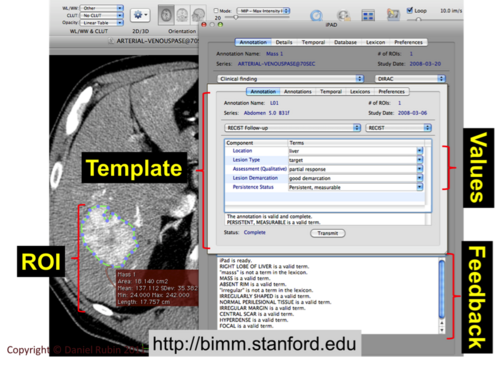
Stanford: Liver lesion
Use case incomplete, verbal description only
- Contact: Daniel Rubin
- Image type: liver CT
- Annotation: N/A
- Markup: uni-dimensional measurement
- Tool used: EPAD
- Preprocessing: none
|
|
NCI: BreastDx
- Contact: Justin Kirby
- Image type: breast MRI
- Annotation: template-based. Template used for textual annotation: File:NCI BreastDx AIM template.xml
- Markup: uni-dimensional measurement of the lesion.
- Tool used: TCGA ClearCanvas
- Preprocessing: none
|
500px
|
MGH: GBM
Use case partially complete, markup not included
- Contact: Jayashree Kalpathy-Cramer
- Images: multi-modal brain MRI
- Annotation: template-based. Template used for textual annotation: File:MGH GBM AIM template.xml
- Markup: binary segmentation of ROI (not included in the use case, not supported by ClearCanvas).
- Tool used for the use case: TCGA ClearCanvas. Other tools used: Slicer, Alice.
- Preprocessing: registration, motion correction, resampling (not included in the provided dataset)
|
500px
|
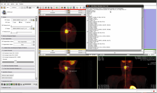
Iowa: Head and Neck cancer
Use case incomplete, verbal description only
- Contact: Milan Sonka
- Images: PET, CT, MRI lungs
- Annotation: yes and no sometimes
- Markup: binary segmentation, unidimensional, bi-dimensional, three-dimensional, 4-dimensional, activity measurement and location
- Tools used: Slicer, Syngo via, Pinnacle, in-house tools
|
|
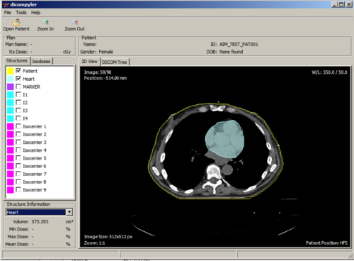
Moffitt: Lung lesion
Use case incomplete, verbal description only. Masks and sample images are available (restricted access)
- Contact: Dmitry Goldgof
- Images: Lung CT
- Annotation: none
- Markup: binary segmentation of ROI
- Tools used: in-house Matlab
|
500px
|
Maastro: Lung lesion
- Contact: Andre Dekker
- Images: Lung CT
- Annotations: N/A
- Markup: DICOM RTSTRUCT
- Tools used: RT-specific tools
|
|
BWH: Prostate cancer
- Contact: Andrey Fedorov
- Images: Prostate MRI
- Annotations: none
- Markup: binary ROI mask drawn on T2w MRI
- Tools used: Slicer
- Preprocessing: registration of the DCE, DWI, T1w parameters to T2w MRI
|
500px
|
OHSU: Breast cancer
Use case incomplete, verbal description only
- Contact: Fred Loney
- Images: DCE MRI
- Annotations: N/A
- Markup: uni- and bi-dimensional measurements, lesion ROI markup
- Tools used: PACS platform, or in-house-developed Matlab-based software
- Preprocessing: N/A


