Projects:RegistrationLibrary:RegLib C03
From NAMIC Wiki
Home < Projects:RegistrationLibrary:RegLib C03updated for v4.1
Back to ARRA main page
Back to Registration main page
Back to Registration Use-case Inventory
updated for v4.1  Slicer Registration Library Case #3: Diffusion Weighted Image Volume: align with structural reference MRI
Input
|
|
|
|
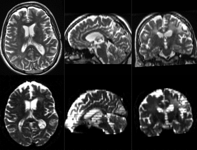
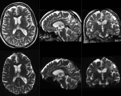
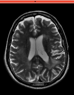
| fixed image/target T2 |
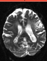
moving image 2a DTI baseline |
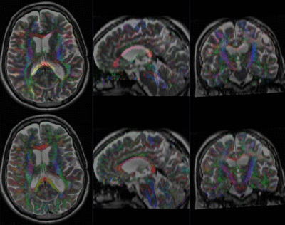
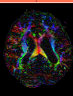
moving image 2b DTI tensor |
Modules
- Slicer 4.1 recommended modules: BrainsFit, Resample DTI Volume, Diffusion Tensor Estimation
- for the Slicer 3.6.3 version of this case see here
Objective / Background
Goal is to align the DTI image with the structural reference T2 scan that provides accuracte anatomical reference.
- Alternate Versions: this example covers the most basic form of directly registering a DTI + baseline to a T2. There is another (more advanced) version that show how to address additional issues of a strong initial rotation and strong voxel-anisotropy for the raw DWI image acquired. You will find the advanced version here.
Download
- Image Data:
- Presets:
Procedure
This assumes you have the following: 1) a T2 reference image, 2) a DTI baseline image and 3) the DTI volume (both obtained from the Diffusion Tensor Estimation module).
- Image Data:
- Overview:
- Using General Registraion (BRAINS), register DTI_baseline to T2 (affine+nonrigid) w/o masking
- Resample the DTI with above transform with the Resample DTI Volume module
- open Registration : General Registration (BRAINS) module
- Input Images: fixed = T2 , moving = DTI_base
- Output Settings:
- Slicer BSpline Transform (create new transform, rename to: "Xf1_DTbase-T2_BSpline")
- Slicer Linear Transform none
- Output Image Volume (create new volume, rename to: "DTIbaseline_Xf1"
- Registration Phases: select/check Rigid , Rigid+Scale, Affine, BSpline
- Main Parameters:
- increase Number Of Samples to 200,000
- set B-Spline Grid Size to 5,5,5
- Leave all other settings at default
- click: Apply; runtime < 1 min.
- Resample DTI
- Open the Resample DTI Volume module (found under: All Modules)
- Input Volume: select DTI
- Output Volume: select create new Diffusion Tensor Volume,and rename it to DTI_Xf2
- Reference Volume: select T2
- Transform Parameters: select transform "Xf2_DTI-T2_masked, Deformation Field: none ; check the displacement checkbox
- Leave all other settings at defaults
- Click Apply; runtime ~ 2 min.
- Go to the Volumes module, select the newly produced DTI_Xf2 volume
- under the Display tab, select Color Orientation from the Scalar Mode menu
- set T2 as background and new DTI_Xf2 volume as foreground
- fade between back- and foreground to see DTI overlay onto the T2 image
Registration Results (click to enlarge)
| baseline & T2 before registration | baseline to T2 after affine+nonrigid alignment | DTI and T2 before & after registration |
Keywords
MRI, brain, head, intra-subject, DTI, DWI
Discussion: Key Strategies
- the two images have identical contrast, hence we could consider "sharper" cost functions, such as NormCorr or MeanSqrd. But because of the strong distortions and lower resolution of the moving image, Mutual Information is recommended as the most robust metric.
- often anatomical labels are available from the reference scan. It would be less work to align the anatomical reference with the DTI, since that would circumvent having to resample the complex tensor data into a new orientation. However the strong distortions are better addressed by registering the other direction, i.e. move the DTI into the anatomical reference space.
- in this example the initial alignment of the two scans is very poor. The strongly oblique orientation of the DWI makes an initial manual alignment step necessary. This step should occur before converting to the DTI to avoid interpolation artifacts.
