2010 Summer Project Week Prostate MRI Segmentation
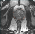
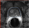
T2w MRI with prostate gland outlined


Key Investigators
- BWH: Andriy Fedorov
- GaTech: Yi Gao
Objective
Segmentation of the prostate gland is one of the steps in the planning of intra-operative guidance for image-guided biopsy and brachytherapy procedures. Accurate segmentation of the gland may be important for accurate calculation of the dose distribution in brachytherapy, and can be critical for accurate registration using surface-based approaches. We would like to automate the segmentation process, to reduce processing time, and improve its accuracy and reproducibility.
We will evaluate the applicability of the prostate segmentation tools developed by Georgia Tech (Yi Gao) on the 1.5T and 3.0T scans with and without endo-rectal coil.
Approach, Plan
This project is a follow-up on the previous developments in projects weeks of 2009-2010 (see references).
Our plan is to:
- make sure the software is compiled and applied correctly
- review processing pipeline
- evaluate the accuracy of segmentation compared with the manual outlines (we will use the publicly available dataset from MICCAI 2009 Prostate segmentation challenge (1.5T) as well as the images from on-going image-guided biopsy program at BWH (3T T2w, Siemens Magnetom)
Progress
Delivery Mechanism
This work will use the prostate segmentation tools earlier contributed to NAMIC Sandbox: http://viewvc.slicer.org/viewcvs.cgi/trunk/Queens/ProstateSeg