Difference between revisions of "2014 Summer Project Week:Cardiac-Congenital"
From NAMIC Wiki
| Line 3: | Line 3: | ||
Image:PW-MIT2014.png|[[2014_Summer_Project_Week#Projects|Projects List]] | Image:PW-MIT2014.png|[[2014_Summer_Project_Week#Projects|Projects List]] | ||
Image:WMH_T1.png|Clinical Stroke Image | Image:WMH_T1.png|Clinical Stroke Image | ||
| − | Image: | + | Image:CongenitalHeartModels.png|Patient-specific heart model |
| − | Image: | + | Image:CongenitalHeartModels2.png|Printed model |
</gallery> | </gallery> | ||
Revision as of 16:06, 23 June 2014
Home < 2014 Summer Project Week:Cardiac-Congenital
Clinical Stroke Image
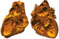
Patient-specific heart model
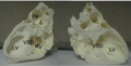
Printed model


Key Investigators
- Danielle Pace, MIT
- Adrian Dalca, MIT
- Polina Golland, MIT
Project Description
This project involves semi-automatic segmentation of gated 3D magnetic resonance images of hearts with congenital heart defects. Our aim is to create patient-specific heart models for surgical planning, which can be viewed either graphically on a computer, or with a 3D printer to create a physical model for surgeons.
We have had initial success in significantly reducing segmentation time with the following pipeline: 1) User manually segments ~10 axial slices 2) Segment the remaining slices using patch-based majority voting.
Objective
- Continue testing the patch based methods on an additional five datasets.
- Address main remaining challenge: segmenting thin interior heart walls.
Approach, Plan
- Manually segment ~10 axial slices for each of the five additional datasets.
- Explore remaining parameters for the patch-based segmentation: e.g. weighted voting, varying k in k-nearest neighbors patch lookup