File list
From NAMIC Wiki
This special page shows all uploaded files.
| Date | Name | Thumbnail | Size | Description | Versions |
|---|---|---|---|---|---|
| 15:16, 7 November 2011 | 20111102 ANNSegmentation.pptx (file) | 5.58 MB | Eun Young Kim's presentation on Artificial Neural Network Segmentation of neural anatomical structures. | 1 | |
| 15:15, 7 November 2011 | ANN Segmentation thesis.pdf (file) | 7.46 MB | Eun Young Kim's thesis on Artificial Neural Network (ANN) segmentation of neural anatomical structures. | 1 | |
| 21:46, 12 October 2011 | NAMIC HD DataDescription.pdf (file) | 2.98 MB | Describes the output of the new autoworkup processing and the DTI processing pipeline. | 1 | |
| 21:42, 12 October 2011 | NAMIC HD DWI DTI.xlsx (file) | 63 KB | Spreadsheet detailing NAMIC_HD shared data anatomical and DWI imaging quality / status. | 1 | |
| 21:24, 7 September 2011 | Anonymized factorsExternalIDs with blage mriDate scanID daysSince.xlsx (file) | 58 KB | 2 | ||
| 21:07, 7 September 2011 | NAMIC+Factors.doc (file) | 43 KB | Explanation of anonymous clinical factors for HD data. | 1 | |
| 14:43, 25 July 2011 | Fetch NAMIC HD data.sh.gz (file) | 1 KB | 4 | ||
| 14:42, 25 July 2011 | Fetch NAMIC HD derived data.sh.gz (file) | 989 bytes | 5 | ||
| 14:05, 24 June 2011 | NAMIC HD Xnat Interface.png (file) |  |
172 KB | Screenshot of xnat interface for NAMIC_HD project. | 1 |
| 02:05, 21 June 2011 | NAMIC DATA SHARING.doc (file) | 31 KB | 1 | ||
| 16:02, 14 January 2011 | DWI TemplateDataRequest.doc (file) | 29 KB | Template of the letter for requesting access to the UIowa DWI Traveling Human Phantom data. | 1 | |
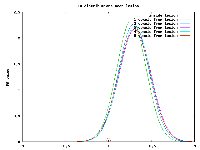
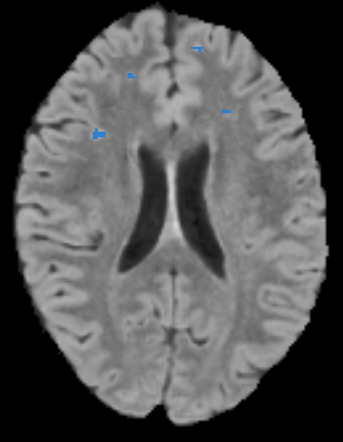
| 14:02, 25 June 2010 | Fa dists.png (file) |  |
5 KB | Distribution of Fractional Anisotropy values for voxels near lesions. | 1 |
| 23:55, 15 June 2010 | LongitudinalLesionComparison TutorialContestSummer2010.zip (file) | 13.48 MB | Data for the Longitudinal Lesion Comparison tutorial for the June 2010 Project Week tutorial contest. | 1 | |
| 18:01, 13 May 2010 | BrainVentriclesAndLesions.png (file) |  |
182 KB | Slice of brain overlaid with ventricles and lesions as volumes | 1 |
| 17:47, 13 May 2010 | CompareViewLDiffZoomedIn.jpg (file) |  |
454 KB | Compare Layout zoomed in with baseline and follow-up FLAIR images overlaid with the lesion difference image where yellow represents lesion in time1 and time2, red is lesion gained in time2, and blue is lesion lost in time2. | 1 |
| 17:43, 13 May 2010 | ComapareViewL1L2Overview.jpg (file) |  |
473 KB | Compare Layout overview of difference between baseline and followup of lupus lesion subject with independent lesion segmentations | 1 |
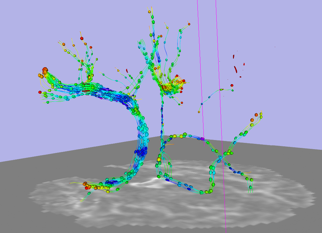
| 15:48, 8 January 2010 | LesionTractsNear.png (file) |  |
346 KB | DTI Tracts near lupus lesions, lines and glyphs, zoomed in. | 1 |
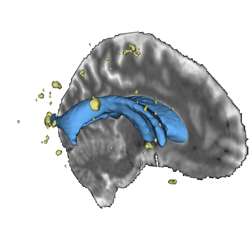
| 15:47, 8 January 2010 | LupusLesionTractsFar.png (file) |  |
218 KB | DTI tracts intersecting lupus lesions | 1 |
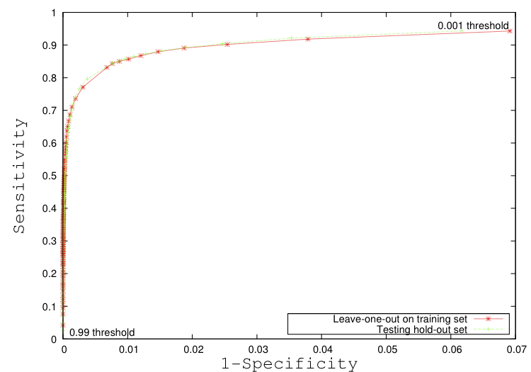
| 21:13, 6 January 2010 | Scully Figure4.png (file) |  |
45 KB | ROC Curve for automated lupus lesion segmentation results training and test sets | 1 |
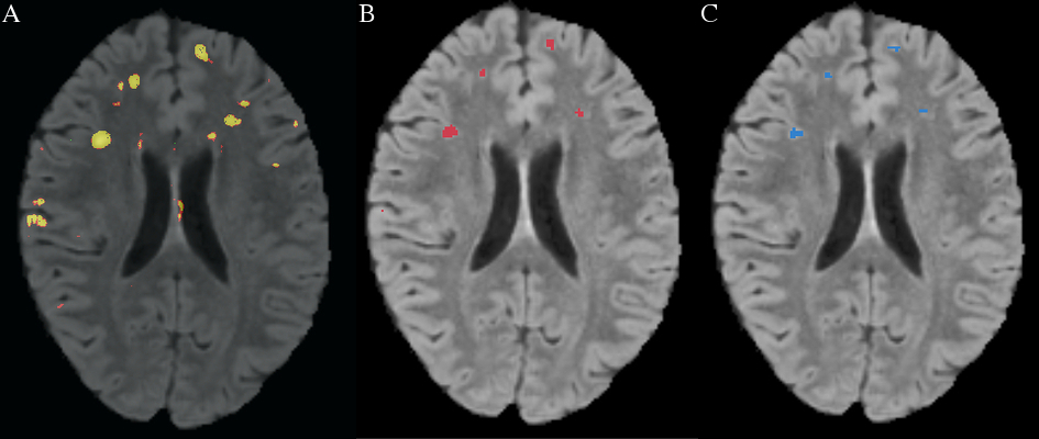
| 21:11, 6 January 2010 | Scully Figure3.jpg (file) |  |
183 KB | Lupus lesion automated segmentation output compared to manual | 1 |
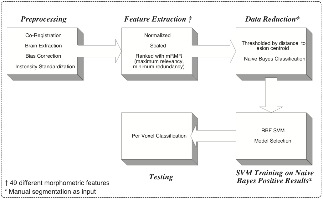
| 21:04, 6 January 2010 | Scully Figure1.png (file) |  |
88 KB | Lupus Lesion Segmentation Method Flowchart | 1 |
| 21:03, 6 January 2010 | Scully Figure1.pdf (file) | 9 KB | Lupus Lesion Segmentation Method Flowchart | 1 | |
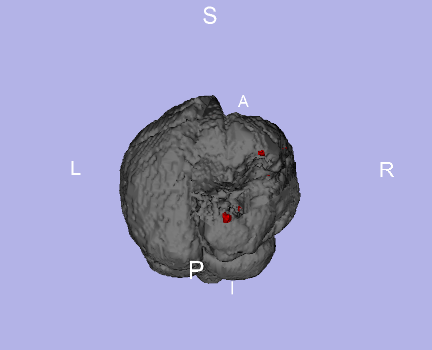
| 13:46, 26 June 2009 | LesionInBrain-PW2009.png (file) |  |
116 KB | Shows lupus lesion model inside brain model. | 1 |
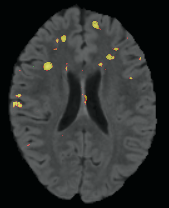
| 17:11, 11 June 2009 | LesionThreshold-PW2009.png (file) |  |
72 KB | Predicted lesion segmentation after thresholding the heat map. (for project week 2009) | 1 |
| 17:10, 11 June 2009 | LesionHeatMap-PW2009.png (file) |  |
90 KB | Predicted lupus lesion heat map for project week 2009 page | 1 |
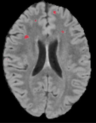
| 17:07, 11 June 2009 | LesionManual-PW2009.png (file) |  |
72 KB | Manual Lupus lesion segmentation for project week 2009 page. | 1 |